Return to Lab 01
Return to Slide List
Benign Neoplasm - Intradermal Nevus (Mole) (PH
2300) (pp. 116-117)
(Be able to
identify this slide; identify normal epidermis and abnormal epidermis;
differentiate between normal epidermis and abnormal epidermis.)
Note that the stratified squamous epithelium of the epidermis has undergone
proliferation. The epidermis is very thick and contains excessive accumulations
of brown melanin. Compare the thickness of the epidermis in the mole with that
of the normal epidermis at the edges of the section. Move the slide back and
forth to do this. Then return the slide to the original position.
Intradermal
nevus (40X1.6)
Normal epidermis (100X2.0)


Dermis (red at bottom), epidermis (blue area and
dark Dermis (red at left),
normal epidermis (dark layer)
layer), air (clear area at right), normal
epidermis (dark
layer in lower region) borders on abnormal thickened
epidermis of nevus (upper blue region with dark surface
layer)
Intradermal nevus (40X1.6)
Intradermal nevus (100X2.0)

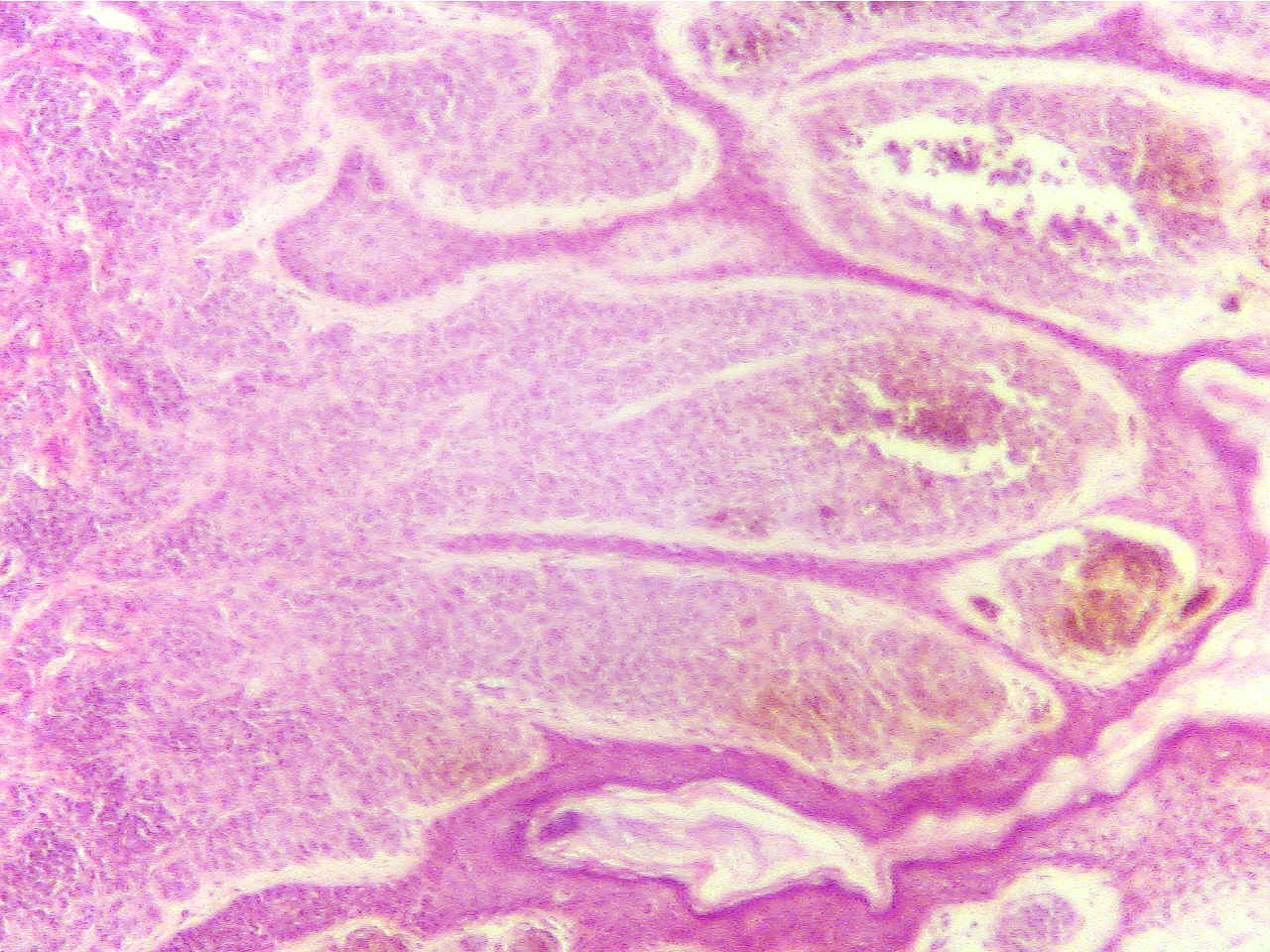
Very thickened and rough epidermis (blue with
dark Very
thickened epidermis (blue with dark surface layer) with
surface layer) and brown melanin masses, dermis
(red) brown melanin masses
at extreme bottom
This neoplasm is considered benign if it does not spread but if it invades
into the dermis it would be considered malignant and called a melanoma.
* To which of the cancer warning signs does this relate?
* Name one important etiological factor in the development of malignant
melanoma.
Return to Lab 01
Return to Slide List
8Copyright
2001 - Augustine G. DiGiovanna - All rights reserved.
This
material may not be reproduced or distributed in any form or by any means, or
stored in any data base or retrieval system without prior written permission is
obtained from Augustine G. DiGiovanna, Ph.D., Professor of Biology,
Salisbury University, Salisbury, MD 21801.