End-Stage Kidney - (Kidney in Diabetes - PH 1765)
(Be able to identify this slide as end stage kidney; identify
normal glomeruli, abnormal glomeruli, tubules, and fibrotic inflammed area; and
differentiate between this slide and slides showing normal kidney.)
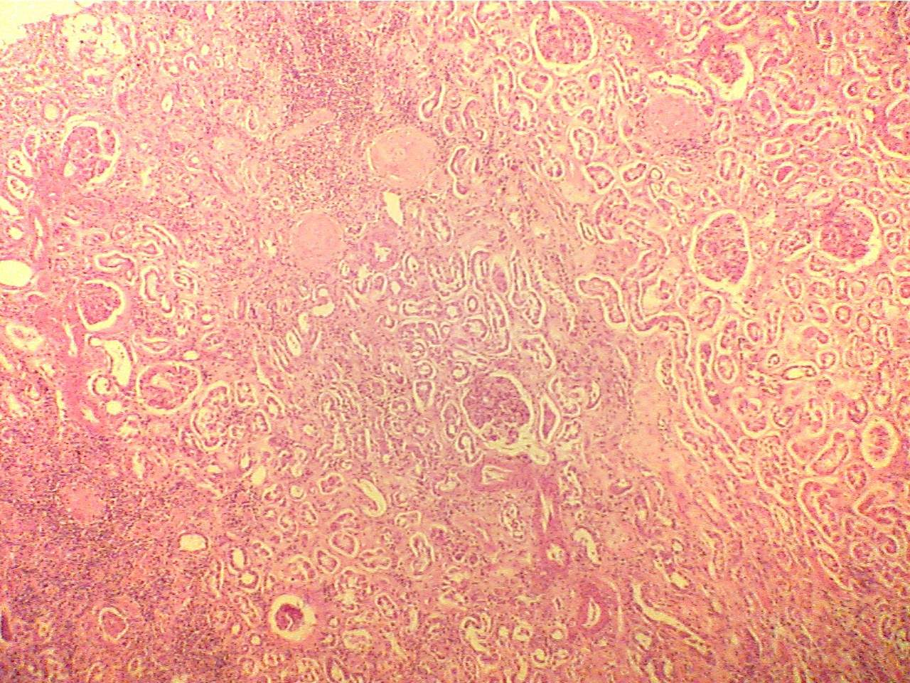
Regardless of the starting point of each specific type of chronic renal
disease, the end is the same, destruction of all parts of the nephron.
This final stage is called end-stage kidney. This particular slide
shows end-stage kidney due to diabetes. (pp. 713-715, Fig. 46-18)
Compare this slide with that of the normal kidney and with Fig. 46-11
on page 706 of the text. Note that both the glomeruli and tubules are abnormal.
Many of the glomeruli are hyalinized (replaced by pink protein material)
and many of the tubules show fibrosis. There is also blood present (RBCs) within
the tubules.
End stage kidney (ESK) cortex
(40X2.0)
End stage kidney (ESK) cortex (100X2.0)
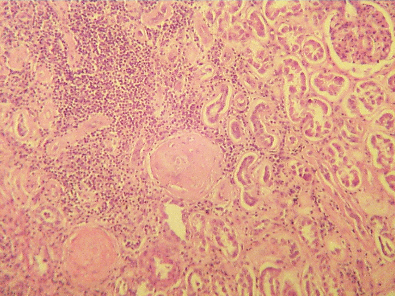
Three normal glomeruli (upper right quadrant),
Normal glomerulus (upper right), two
two hyalinized glomeruli (above center), few
tubules, hyalinized glomeruli (center, lower left),
concentrations of inflammatory cells
(lower
concentration of inflammatory cells (upper left quadrant)
left, upper middle)
End stage kidney (ESK) cortex (100X2.0)
End stage kidney (ESK) glomerulus (320)
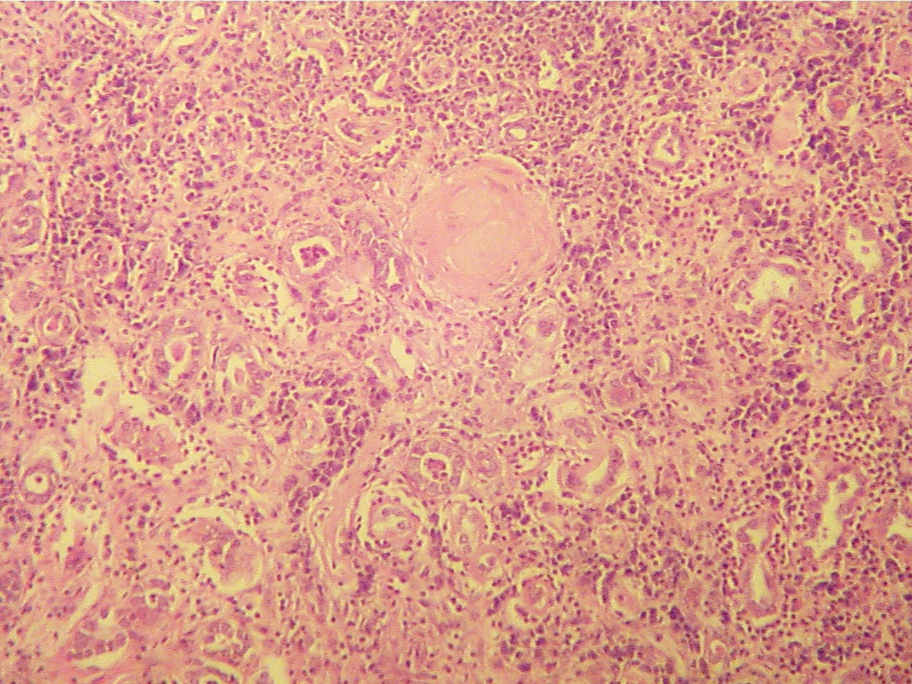
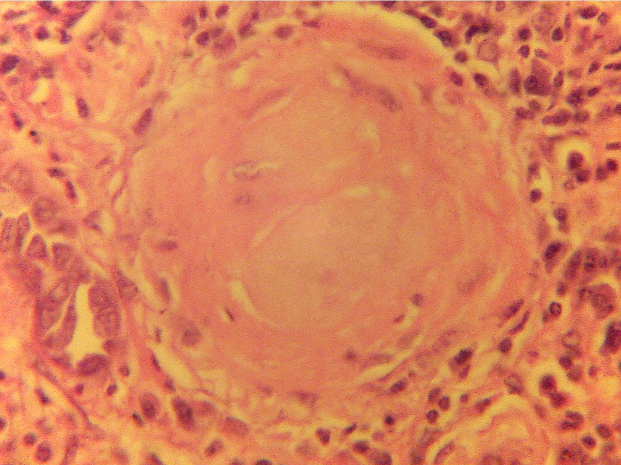
Hyalinized glomerulus (above center), few tubules Hyalinized glomerulus (mostly solid pink with few
around glomerulus and throughout, inflammatory cells
dark blue nuclei) surrounded by small dark blue
and fibrosis (areas between structures).
inflammatory cells rather than tubules.
43.
End stage renal disease, gross
44.
End stage renal disease, microscopic *