Return to Lab 01
Return to Slide List
Abscess - Chronic Subcutaneous Abscess (PH 2335) (pp. 49-50)
(Be able to identify this slide at 40X or 100X and differentiate between the normal and
abnormal regions.)
Notice the spherical mass of unorganized cells and cell debris surrounded by
a capsule of fibrous connective tissue within the dermis of the skin. Compare
its appearance with the normal dermis by moving the slide back and forth.
Observe the abscess with both the compound and dissecting microscopes and then
return slide to original position.
Subcutaneous abscess (40X1.0)
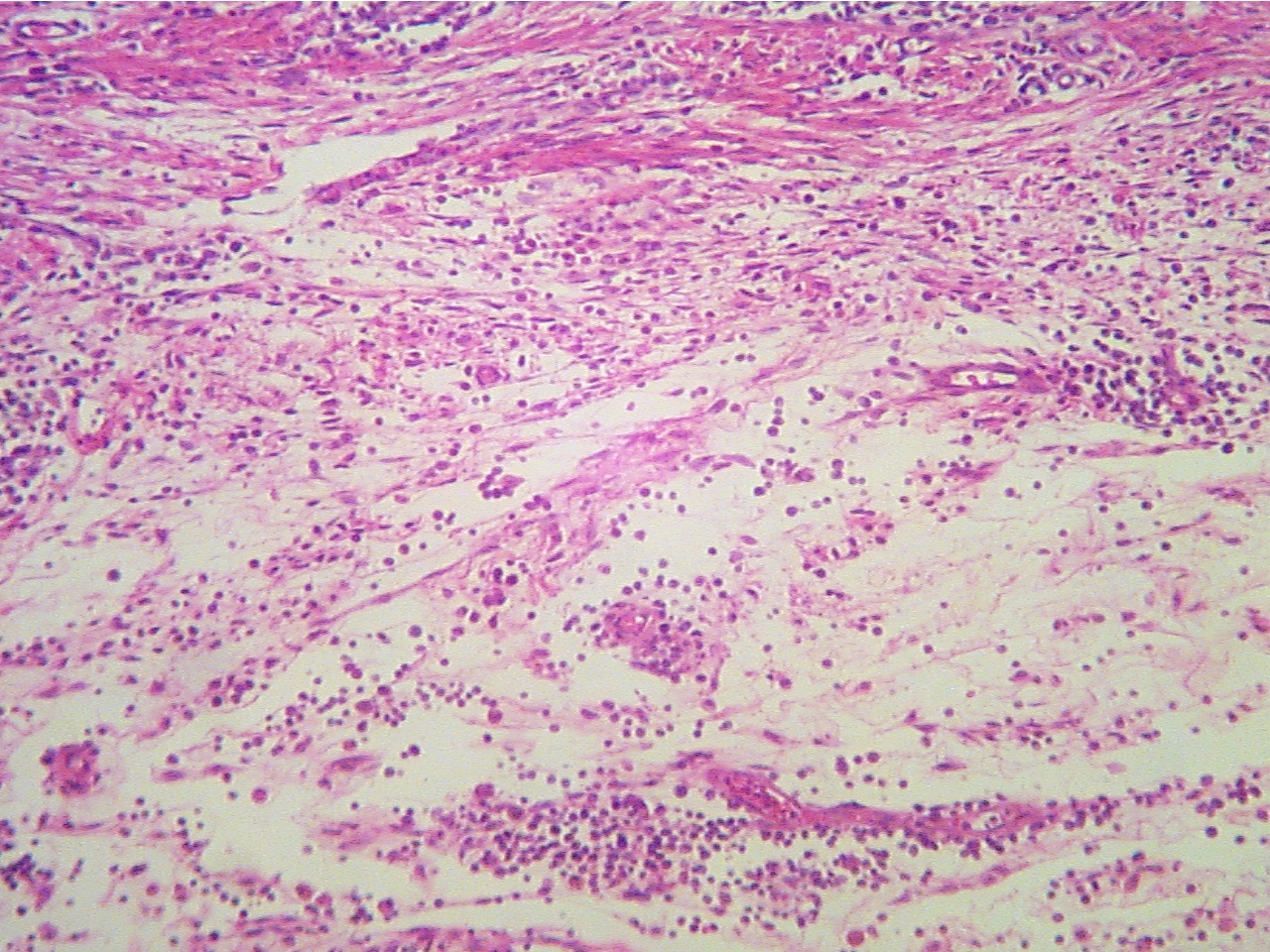
Subcutaneous abscess (100X2.0)
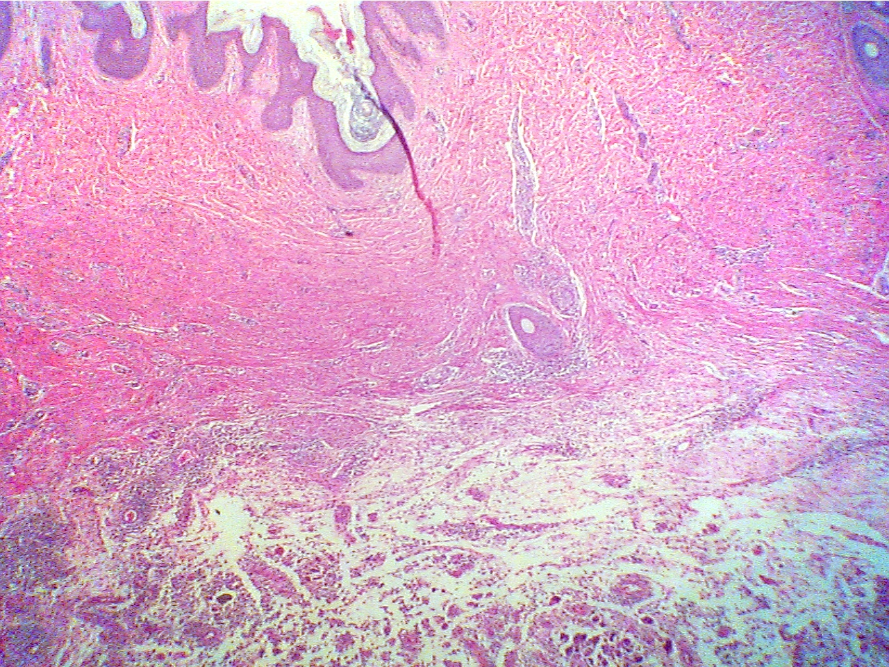
Epidermis (thin dark layer at top), dermis
(thick
Edge of dermis (fibers at top), pus (scattered cells
red layer), edge of abscess (scattered dark
cells
and debris in clear liquid at bottom)
and debris at bottom)
Subcutaneous abscess (100X2.8)
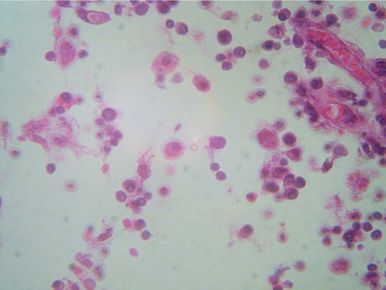
Subcutaneous abscess (400X2.0)
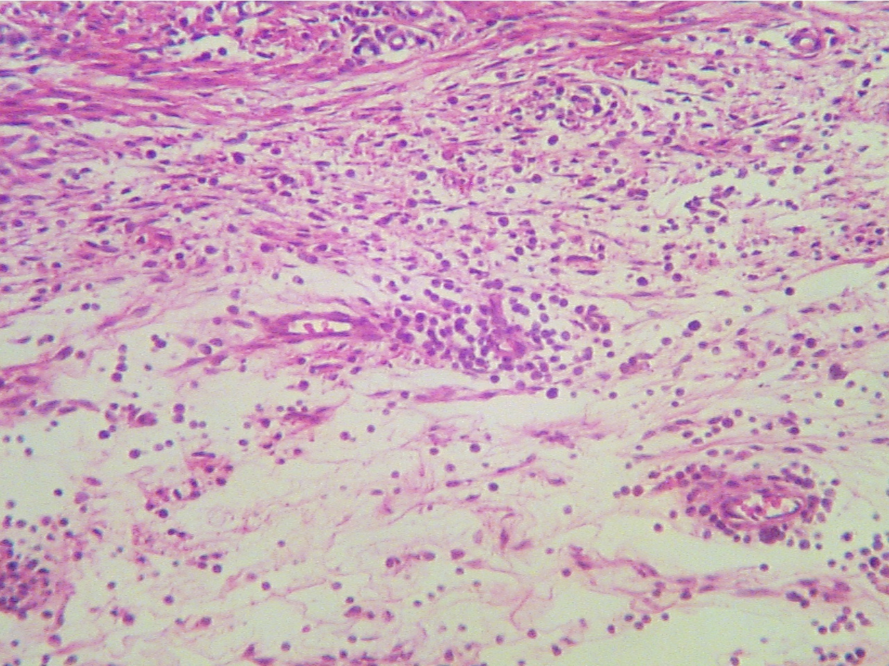
Edge
of dermis (fibers at top), pus (scattered cells
and Pus (scattered inflammatory cells,
tissue debris,
debris in clear liquid at
bottom)
liquid (clear), remains of two capillaries (right)
* What is the name of the
material filling the abscess?
Return to Lab 01
Return to Slide List
8Copyright
2001 - Augustine G. DiGiovanna - All rights reserved.
This
material may not be reproduced or distributed in any form or by any means, or
stored in any data base or retrieval system without prior written permission is
obtained from Augustine G. DiGiovanna, Ph.D., Professor of Biology,
Salisbury University, Salisbury, MD 21801.