Early lesion (40X1.0 - a1)
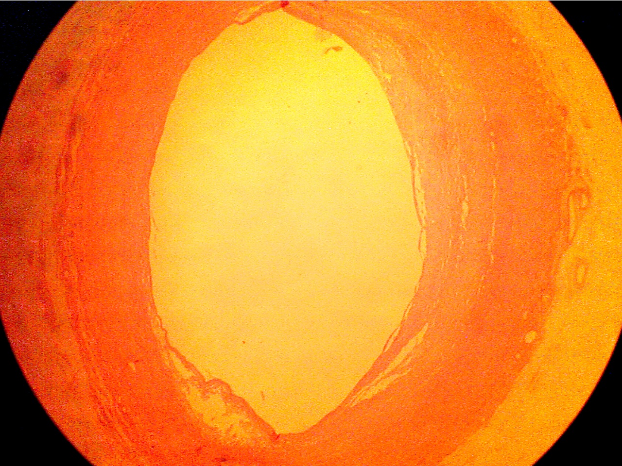
Normal artery wall at left, lumen in center,
lesion (thickened wall) at right.
Normal region (100X2.0 - a1) Early lesion (100X2.0 - a2)
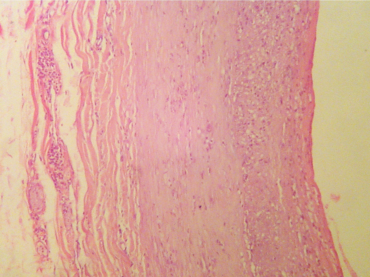
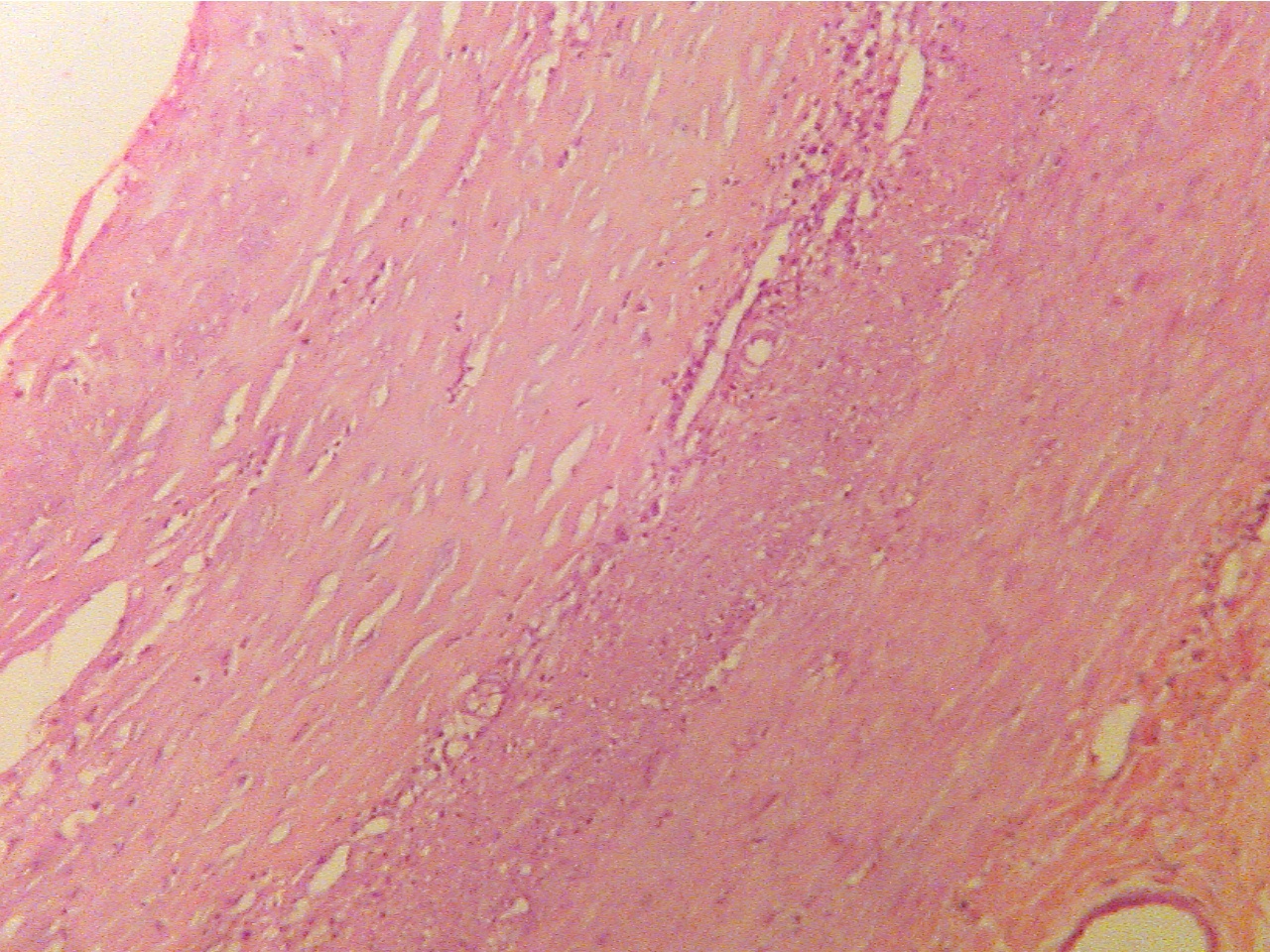
Layers are areolar tissue (left), externa (thick pink Lumen at upper left, endothelium (thin layer),
collagen fibers), smooth muscle (thick middle layer), intima, fibrous plaque within intima, intima (darker granular
tunica intima (thick granular layer), endothelium layer), smooth muscle (thick pink), externa (lower right)
(thin red layer), lumen at right
Advanced lesion (40X1.0 - b1)
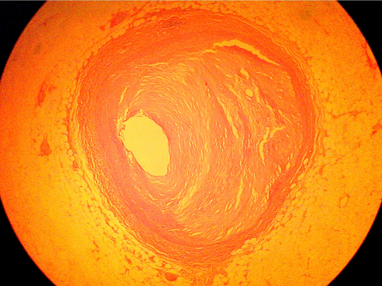
Lumen at left of center, lesion at right
Normal region (100X2.0 - b1) Advanced lesion (100X2.0 - b2)
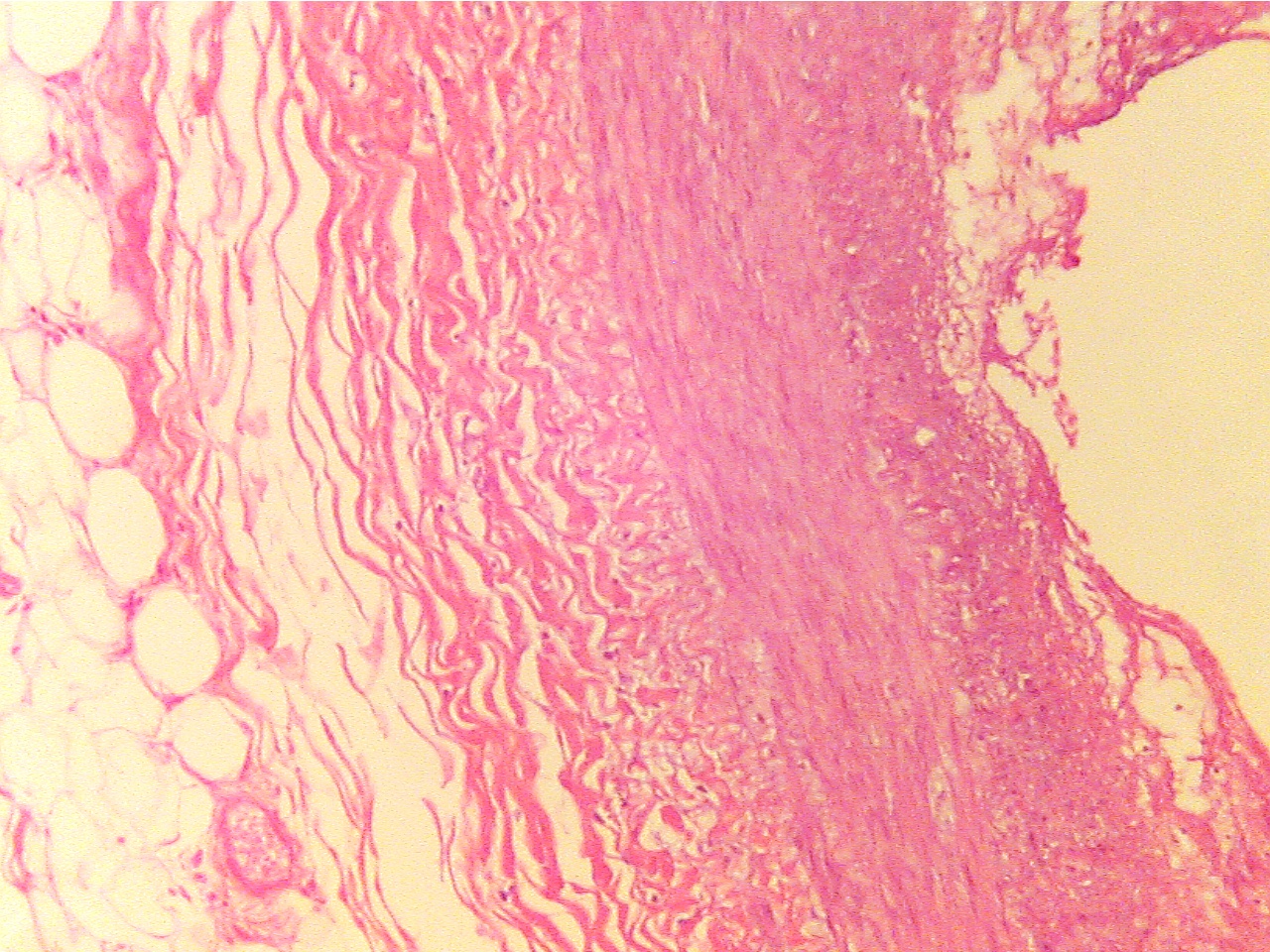
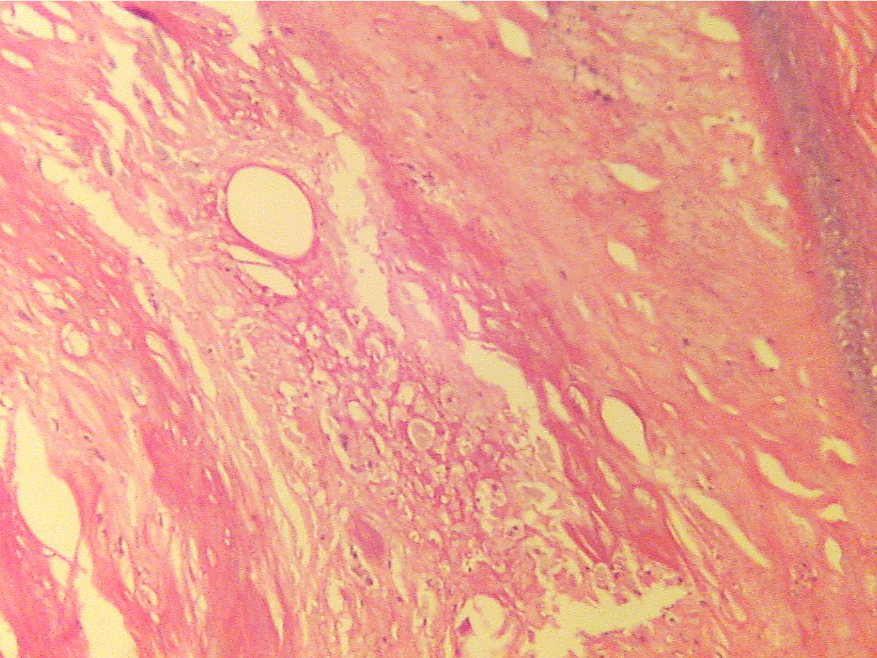
Externa at left, lumen at right Lumen at far lower left corner, lesion of mixed
substances and cells fills center region
Occlusive lesion (40X1.0 - c1)
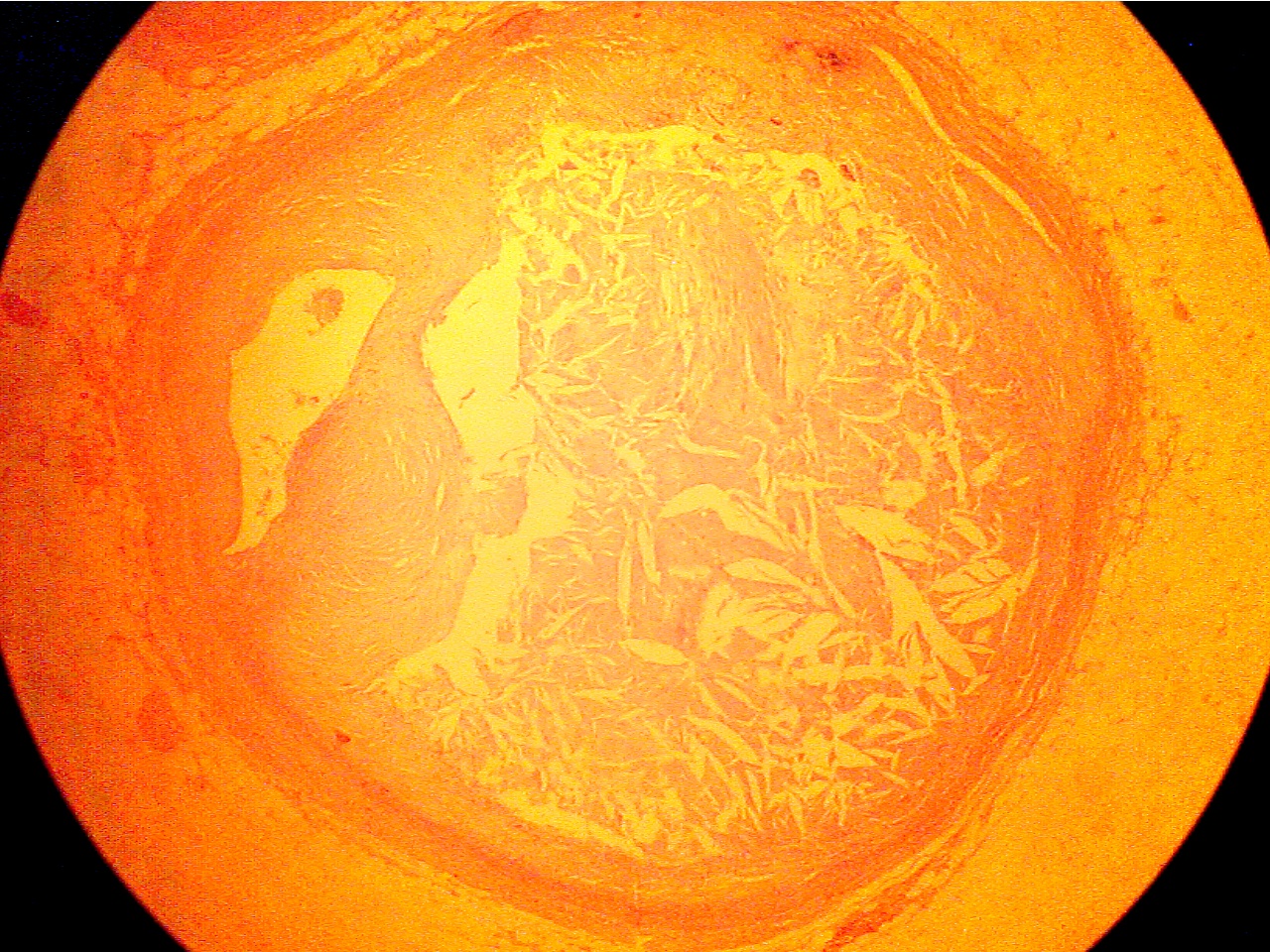
Lumen at far left of center, crystalline lesion fills center
Fibrous plaque (100X2.0 - c1) Plaque replaces intima and smooth muscle (100X2.0 - c2)
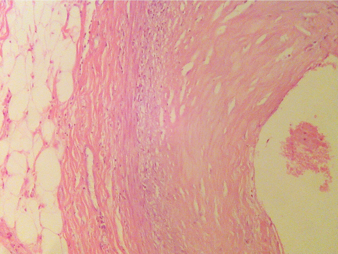
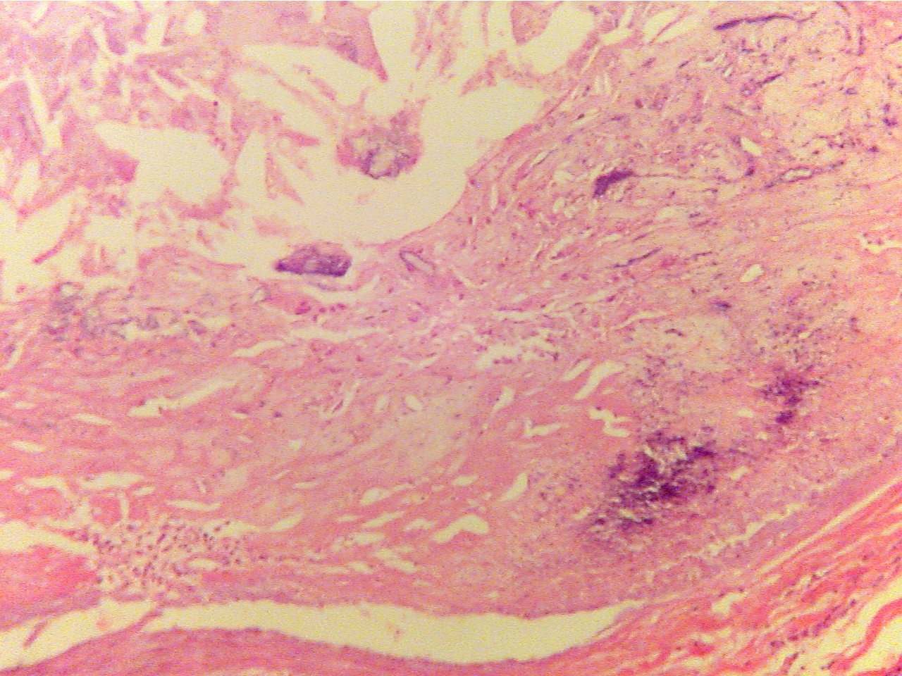
Layers are areolar tissue (left), externa (red collagen Amorphous acellular material (upper left) and cellular
fibers), smooth muscle (thin red middle layer), tunica and fibrous plaque (center) replace intima and smooth muscle,
intima (blue granular layer), fibrous plaque (thick, pink), externa (collagen fibers) remains at lower right
endothelium (thin layer along lumen), lumen (at right)
Center of plaque (100X2.0 - c3) Endothelium (400X2.0 - c4)
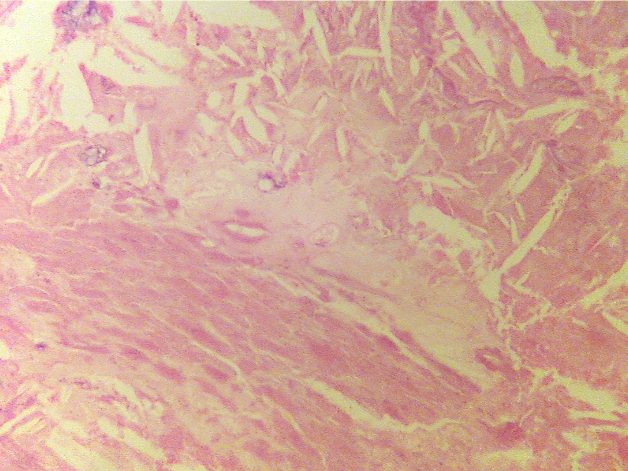
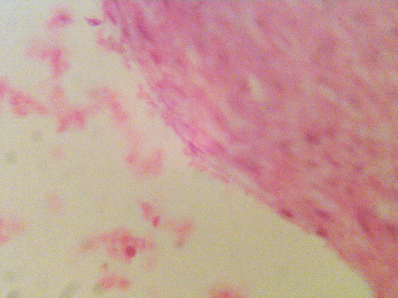
Center of amorphous acellular region Normal endothelium (lower right bordering lumen), plaque
of plaque, much of which appears crystalline (right, underlies endothelium), lumen (left), damaged
endothelium with adhering RBCs (middle and top)
* Clinically significant lesions (which produce myocardial ischemia) usually obstruct over percent of the vessel lumen. (p. 461)