Return to Lab 01
Return to Slide List
Adenocarcinoma - Adenocarcinoma of kidney (PH 1720) (pp.
114-116)
(Be
able to identify the slide showing kidney cancer; identify normal glomeruli and
tubules; and differentiate between normal and diseased regions.)
Note that tubular cells have undergone extensive proliferation and have lost
their characteristic appearance. They look more like undifferentiated cells and
there is extensive stroma filled with red blood cells between the cancer cells.
See Fig. 8-9. This situation is very different from that of the benign adenoma
of the colon. Compare the tumor with the normal kidney structure (tubules and
glomeruli) by moving the slide back and forth. Note that the tumor is displacing
and invading into the normal kidney tissue. There is no sharp boundary between
normal and malignant tissue. Return the slide to the area of the tumor.
Normal kidney - cortex region(40X2.0)
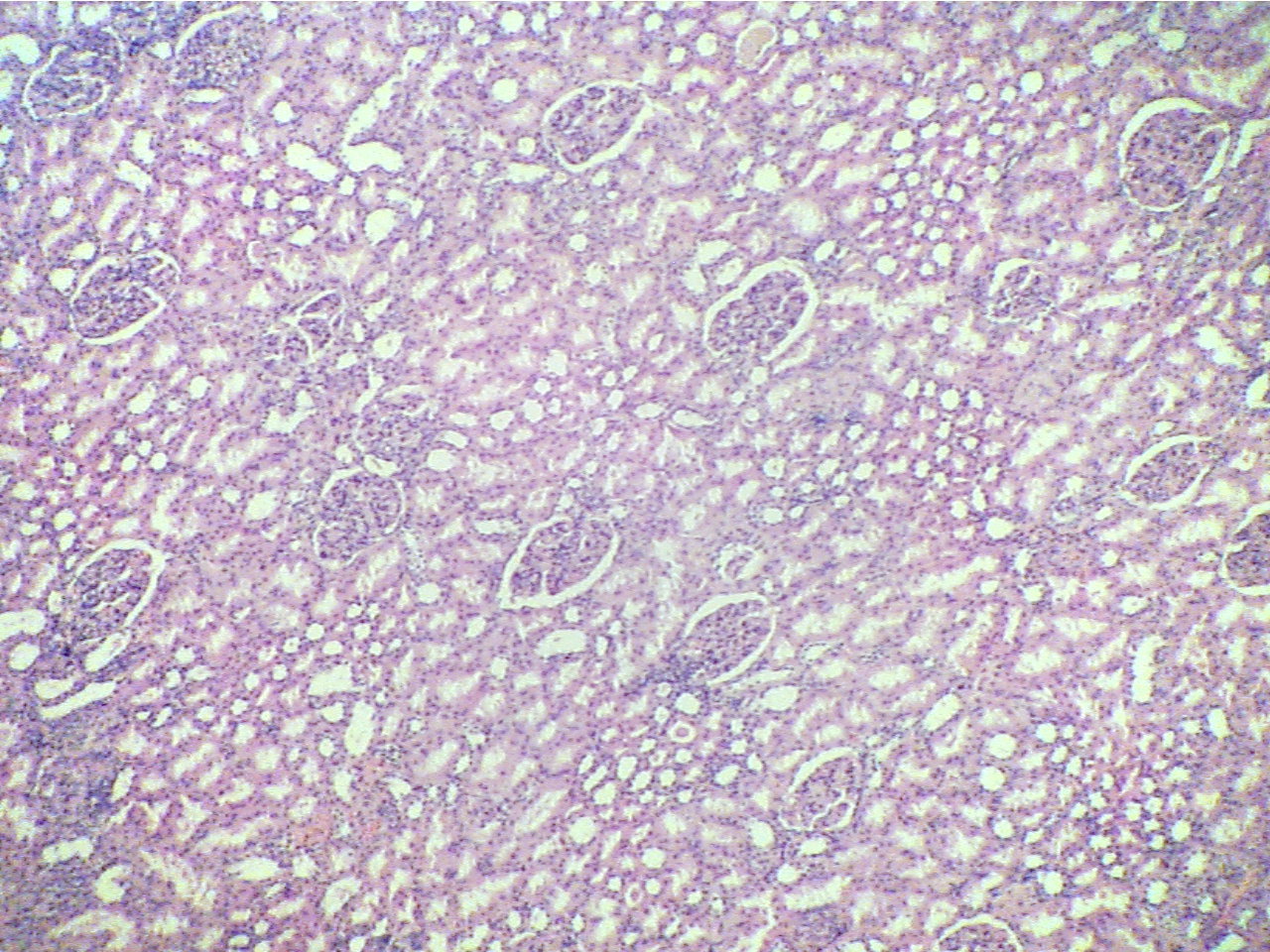
Approximately twelve glomeruli, each surrounded by tubules
Adenocarcinoma of kidney (40X2.0)
Adenocarcinoma of kidney
(40X2.0)
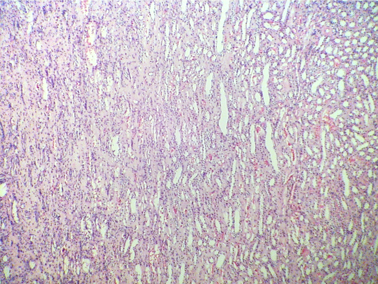

Normal kidney tubules at right, neoplasia at
left
Neoplasia at right, hemorrhaging neoplasia at center and left
* Why is this called an adenocarcinoma?
Return to Lab 01
Return to Slide List
8Copyright
2001 - Augustine G. DiGiovanna - All rights reserved.
This
material may not be reproduced or distributed in any form or by any means, or
stored in any data base or retrieval system without prior written permission is
obtained from Augustine G. DiGiovanna, Ph.D., Professor of Biology,
Salisbury University, Salisbury, MD 21801.