Normal liver (40X2.0) Normal liver (400X2.0)
Return to Lab 01
Return to Lab 10
Return to Slide List
Development of Cirrhosis of the Liver - Normal Liver (PH 8145),
Fatty Metamorphosis (PH 1525) and Cirrhosis of the Liver (PH 1520) (pp. 30-31,
389)
(Be able to identify
slides of normal liver, fatty liver, and cirrhosis; identify normal liver cells,
fatty liver cells, and steaks of fibrotic (scar) tissue; and differentiate
between slides showing normal and abnormal liver.)
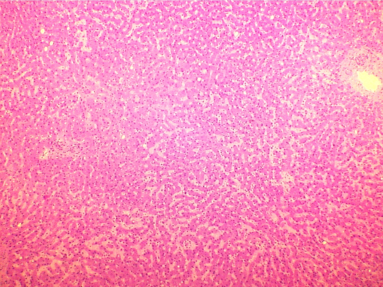
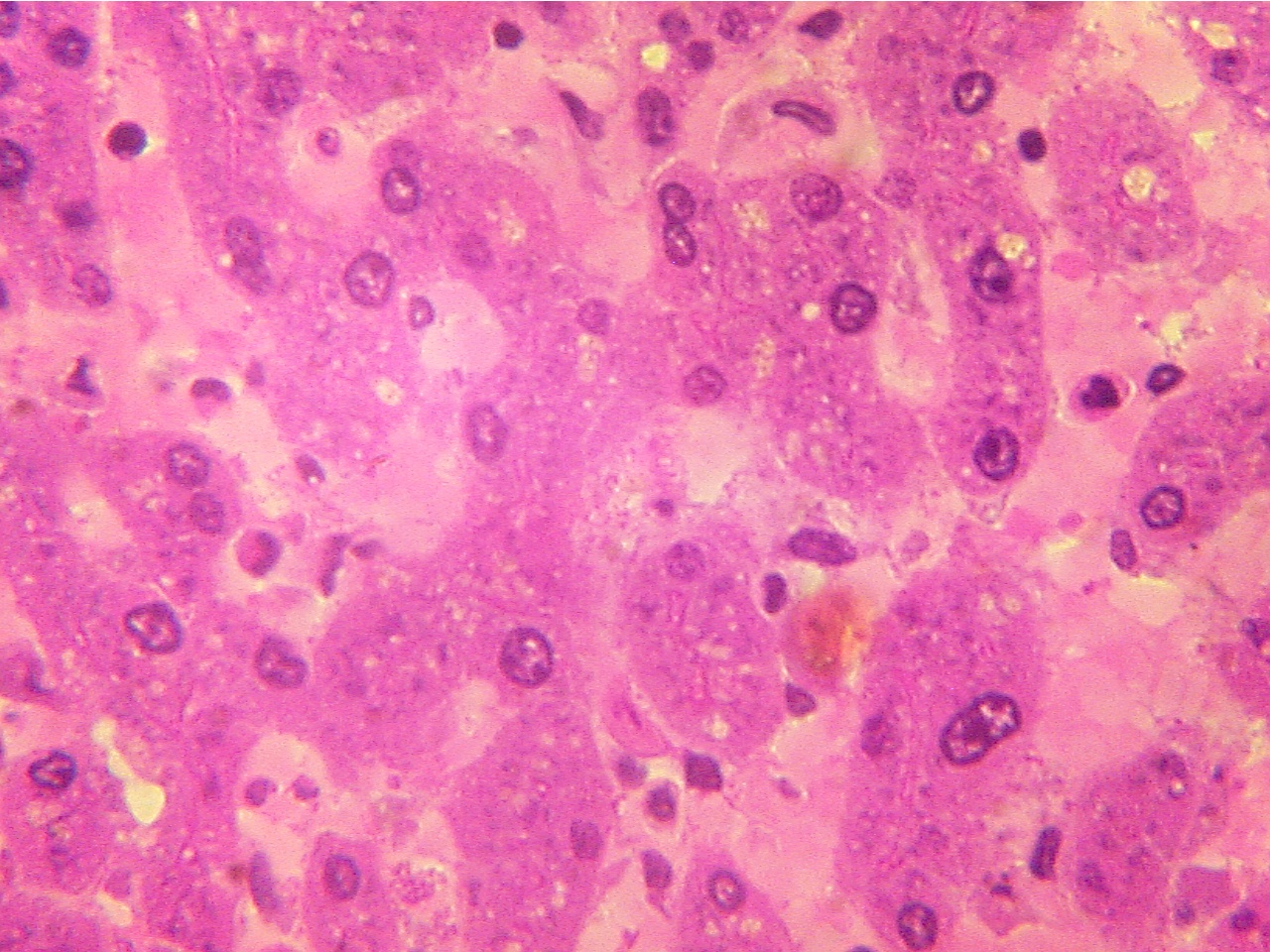
First study the slide of normal liver. Notice the size and shape of the liver
cells, the purple nuclei and pink cytoplasm. Notice the pale colored sinusoids (specialized
capillaries) situated between strands of liver cells.
Normal liver
(40X2.0)
Normal liver (400X2.0)
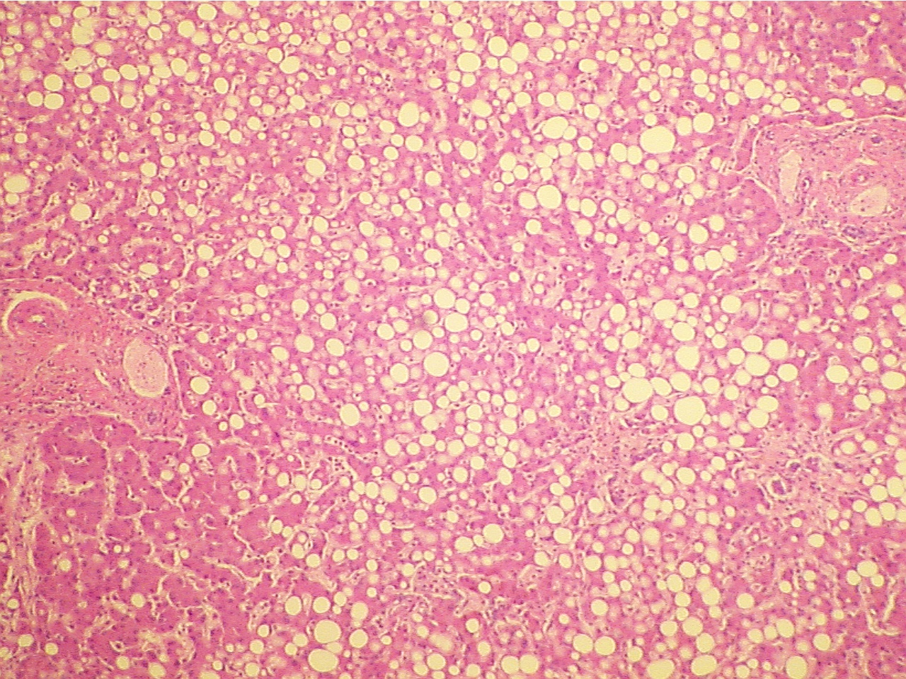
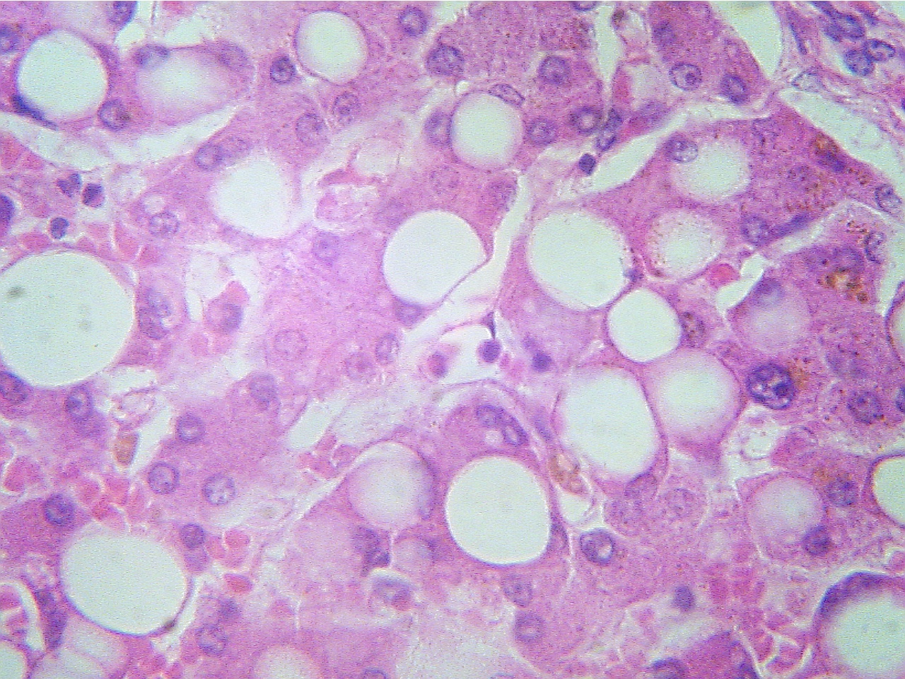
On the slide of fatty degeneration (fatty
metamorphosis), you will note large clear bubbles or vacuoles within the
liver cells. Notice how these vacuoles occupy a large proportion of the cell's
volume and push the cytoplasm up against the cell membrane, forming "signet ring
cells". These vacuoles contain fat which has accumulated due to abnormal
biochemical reactions within the cell. These changes occur with various types of
injury to the liver, including the early stages of cirrhosis.
Fatty liver
(40X2.0)
Fatty liver (400X2.0)
* Is fatty degeneration a reversible or irreversible change?
With continued assault to liver cells, cells will die and be replaced by scar
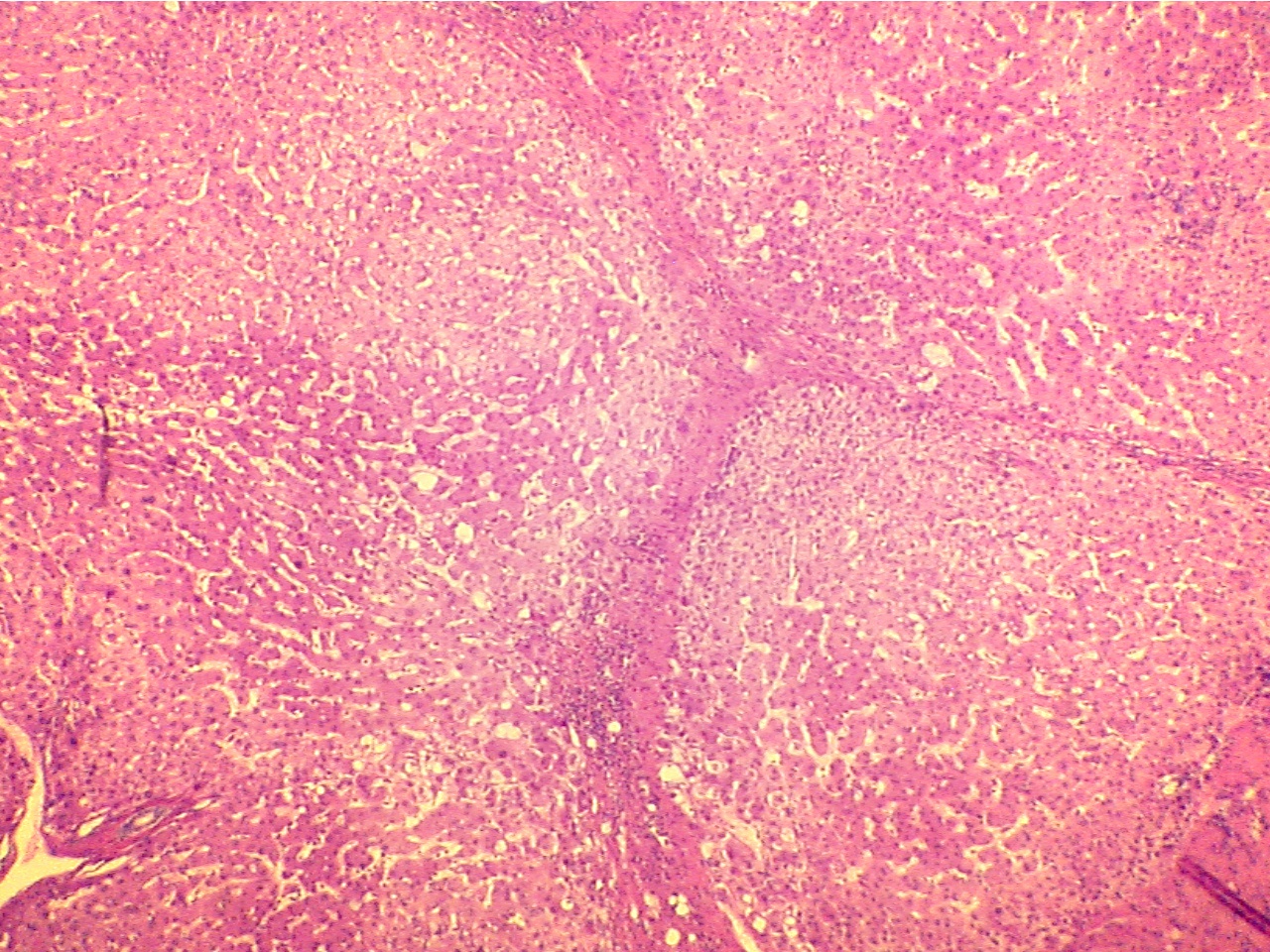
tissue. On the slide of cirrhosis of the liver, notice bands of fibrous
scar tissue which extend through the liver and separate lobules of functioning
liver cells. The many small blue cells are inflammatory cells, which are WBCs
that invaded the area and indicate that inflammation is occurring. (See also the photograph of Liver/Cirrhosis).
Cirrhosis
(40X2.0)
Cirrhosis (400X2.0)
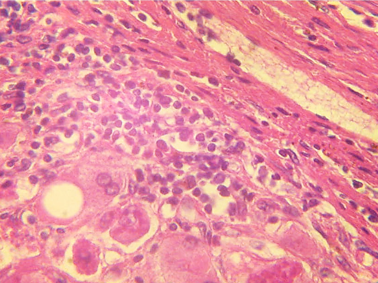
Fibrosis tissue (top) and inflammatory cells (middle)
* What is a common cause of cirrhosis?
* Is cirrhosis reversible or irreversible?
Return to Lab 10
Return to Slide List
8Copyright
2001 - Augustine G. DiGiovanna - All rights reserved.