- can be changed by dilation
Normal cardiac muscle, medium power microscopic *
Heart, dilated cardiomyopathy, gross [XRAY]
Heart, dilated cardiomyopathy, gross
Dr. D.'s Overhead Lecture Notes
Section 2 - REPLACE PAGE NUMBERS WITH PAGES FROM SIXTH EDITION2
Do Not Print This Page
These notes are in your course booklet and in Sec 02-w.htm.
Abnormal accumulation of fluids
A. Congestion = hyperemia
- excess blood in vessels
1. Active congestion = active
hyperemia
- arteries/arterioles
dilate -> excess delivery of blood
- causes
- inflammation, hypoxia, pH, temp., emotions
- effects
= adaptive
Active hyperemia - due to a usually beneficial increased input of
blood from arteriolar and capillary dilation
Example -
for inflammation
1.
Erythema, gross
2.
Erythema and edema, gross
Example - for temperature regulation by integumentary vasodilation
17.Hyperthermia,
scene
Harmful example - edema blocks airway
13.
Edema in larynx, gross
2. acute passive congestion
- local
- blockage of veins
- reduced drainage
of area
- e.g. tumors, posture, clothing
- systemic
- reduced heart function ( cardiac output)
- reduced drainage of veins
- e.g. heart attack, valve disease
- effects
= detrimental (1, 2, & 3)
1. slow flow -> decr.
O2,
incr. CO2,
decr.
nutrients,
incr. wastes -> cell
injury/cell death -> pain and decr.
function
2. slow flow -> thrombosis(Fig.
7-4 p.98)
3. incr. volume -> incr.
pressure -> edema
Note: acute
= short term -> no lasting effects
3. chronic passive congestion
- local
- blockage of veins
- systemic
- decr. heart function
- effects
= detrimental (1, 2, & 3)
1. slow flow -> pain, decr.function
2. slow flow -> thrombosis
3. incr. volume -> edema,
varicose
veins
Note: chronic = long term -> lasting
effects
Examples - liver
3.
Normal liver, cut surface, gross
Note the color and the smooth homogeneous structure of the brown liver
tissue.
55.
Chronic passive congestion (nutmeg liver), gross
Note the spotty color and the uneven structure of the liver tissue.
4.
Normal liver zones, microscopic*
The liver cells make up the prominent pink strands of material. Each liver
cell can be identified by its small round blue nucleus. The more open streaks
between the strands of liver cells are large capillaries (i.e., liver sinusoids)
that contain some blood cells. The large vessels in the lower left region
carry blood to the liver sinusoids, and the large vessel in the upper right
drains blood from the sinusoids. Note that the liver cell cytoplasm appears
solid in color and that most of the liver consists of liver cells and sinusoids.
56.
Chronic passive congestion, liver, microscopic*
57.
Centrilobular necrosis, liver, microscopic*
58.
Chronic passive congestion with "cardiac cirrhosis", liver, microscopic*
Example - spleen
22.
Splenomegaly with portal hypertension, gross
Portal hypertension means high blood pressure in the hepatic portal veins.
These veins carry blood to the liver from the spleen, the pancreas, and
much of the GI tract between the esophagus and the anus. The high pressure
in the portal veins develops because of passive congestion. The passive
congetoins develops because fibrosis in a liver with cirrhosis blocks liver
vessels, preventing blood in the spleen and digestive system organs from
going to the liver.
Effects on veins = varicose
veins = permanently and excessively dilated veins
Examples - esophagus
1.
Normal esophagus, gross
12.
Esophageal varices, gross
Examples - hemorrhoids
118.
Prolapsed true hemorrhoids, gross
Examples - skin
20.
Caput medusae of skin with portal hypertension, gross
B. edema = excess fluid between
cells
i.e., incr.
interstitial fluid
11.
Edema, gross
2.
Erythema and edema, gross
31.
Anaphylaxis with acute laryngeal edema, gross
Examples in lungs - pulmonary edema
4.
Normal lung, microscopic*
Note the thin membranes and open air spaces
5.
Lung, edema, microscopic*
This is at high magnification, note the thick membranes and filled air
spaces
- mechanisms
(1, 2, or 3)
1. incr. BP (hydrostatic pressure)
(e.g. inflammation, hypertension, passive congestion)
2. decr. COP (colloidal osmotic pressure)
(e.g. inflammation, malnutrition, proteinuria, cirrhosis)
3. blocked lymphatics
(e.g. cancer, surgery, infection)
- effects
1. discomfort
2. in lungs = pulmonary edema (p.726)
(sketch)
yields
|
|
|
|
| thicker membrane + filling alveoli
|
|
|
|
|
|
|
|
|
|
|
4.
Normal lung, microscopic*
Note the thin membranes and open air spaces
5.
Lung, edema, microscopic*
This is at high magnification, note the thick membranes and filled air
spaces
6.
Lung, passive congestion, microscopic*
This is at high magnification, note the thick membranes and filled air
spaces
14.
Pleural effusion, serous, gross [XRAY]
Fluids has leaked into the pleural cavity
115.
Lung, interstitial fibrosis, microscopic*
Chronic pulmonary edema can cause pulmonary fibrosis.
3.
in brain -> increased
pressure -> a or b
a. damages neurons
b. compresses vessels -> decr.
flow
Hemorrhage = blood leaks from vessels
- general causes (1,
2, & 3)
1. trauma
to vessel (e.g. cut, crush)
2. disease
weakens vessel
3. low
clotting ability
Normal
peripheral blood, segmented neutrophil and band neutrophil, high power
microscopic
The small blue specks are platelets. Substances released by platelets start
the clotting process.
39.
Hematoma, toenail, gross
A hematoma is a blood clot outside blood vessels or the heart but enclosed
in the body.
37.
Petechial hemorrhages, epicardium, gross
38.
Ecchymoses, skin of arm, gross
34.
Hemorrhagic pulmonary infarction, gross
35.
Hemorrhagic pulmonary infarction, gross
- local effects (1
or 2) (sketch)
1. Hb injures
cells
2. volume disrupts
function
127.
Hemothorax, gross
This hematoma inhibits filling of the lungs during inspiration. It caused
the lungs to collapse (atelectasis).
20.
Hemopericardium, gross
This hematoma around the heart inhibits filling of the heart during diastole.
- systemic effects
(1 or 2)
1. rapid massive
leak -> shock
2. slow chronic
leak -> anemia
- fates of hemorrhaged blood
(1, 2, or 3)
1. peel off (e.g.,
scab)
2. removal of
hematoma
(ex. "black + blue" mark fades)
3. organization
of hematoma (ex. scar formation replaces hematoma)
Blood clot in vessel = thrombus
Normal
peripheral blood, segmented neutrophil and band neutrophil, high power
microscopic
The small blue specks are platelets. Substances released by platelets start
the clotting process.
36.
Platelet, electron micrograph *
33.
Recent thrombus with lines of Zahn, microscopic *
7.
Severe coronary atherosclerosis, gross
9.
Coronary thrombosis, gross
40.
Disseminated intravascular coagulopathy (DIC), fibrin thrombus in lung,
high power microscopic
*
general causes (1, 2, &
3) (sketch)
1. rough lining
of vessel or heart
- breaks platelets -> clotting
2. slow flow
(p.
98, Fig. 7-4)
- clotting factors build up
3. chemical imbalance
- breaks platelets -> clotting
effects (1 or 2)
1. block
vessel at site (p. 100)
2. block
vessel at other site (sketch)
- embolus - embolize
10.
Coronary thrombosis, gross
11.
Coronary thrombosis, microscopic *
Renal
vein thrombosis, gross
Hyaline
thrombus in glomerulus with thrombotic thrombocytopenic purpura (TTP),
microscopic *
fates of thrombi (1 or 2)
1. dissolved
2. organized
78.
Lung, remote organized pulmonary artery embolus, gross
6.
Coronary atherosclerosis with thrombosis, microscopic *
types of emboli
1. thrombi
Thromboembolus - moving blood clot in a blood vessel
Pulmonary embolus
27.
GIF animation of pulmonary thromboembolus, diagram
28.
Pulmonary thromboembolus, gross
29.
Pulmonary thromboembolus, gross
30.
Pulmonary thromboembolus, gross
31.
Pulmonary thromboembolus, microscopic *
32.
Pulmonary thromboembolus, microscopic *
27.
GIF animation of pulmonary thromboembolus, diagram
69.
Lung, pulmonary thromboembolus, gross
70.
Lung, pulmonary thromboembolus, gross
73.
Lung, pulmonary thromboembolus, gross
74.
Lung, pulmonary thromboembolus, low power microscopic*
78.
Lung, remote organized pulmonary artery embolus, gross
2. gas
bubbles
3. plaque
(from atherosclerosis)
4. fat
droplets (from trauma)
1.
Fat embolism, lung, high power microscopic *
2.
Fat embolism, lung, Oil red O stain, high power microscopic *
3.
Fat embolism, glomerulus, high power microscopic *
4.
Fat embolism, brain, gross
5.
Fat embolism, brain, gross
6.
Fat embolism, brain, high power microscopic *
5. cancer
cells (from metastasis)
44.
Metastatic adenocarcinoma, liver, gross [CT]
45.
Metastatic adenocarcinoma, liver, gross
46.
Metastatic adenocarcinoma, liver, gross [CT]
47.
Metastatic adenocarcinoma, liver, microscopic*
effects of emboli
- block
vessels
Diseases of vessel walls - arteries
Arteriosclerosis = "arterial
scarring"
Atherosclerosis = arteriosclerosis
of large arteries
contributing factors = risk
factors (p. 463)
** 1. smoking
** 2. high
blood pressure = hypertension
** 3. high
blood LDLs ~ high blood cholesterol
* 4. genetic predisposition = runs in families
* 5. diabetes mellitus
6. being male
7. age
8. obesity
9. high fat diet
10. sedentary lifestyle
11. stress
12. "the pill" + smoking
13.
pathogenesis (p.
462)
cause(s) ->
deposits in tunica intima -> (1 and 2)
(sketch)
1. inward growth of plaque -> (a and b)
(sketch)
a. narrowness -> *low
blood flow (p.
103)
1.
Normal coronary artery, microscopic *
2.
Mild coronary atherosclerosis, microscopic *
3.
Severe calcific coronary atherosclerosis, microscopic *
4.
Coronary atherosclerosis, cross sections, gross
9.
Coronary thrombosis, gross
10.
Coronary thrombosis, gross
11.
Coronary thrombosis, microscopic *
Left
anterior descending coronary artery, advanced atherosclerosis, gross.
b. roughness -> thrombus ->
*low blood flow (p. 100, p. 102, p. 103)
(sketch)
9.
Coronary thrombosis, gross
10.
Coronary thrombosis, gross
11.
Coronary thrombosis, microscopic
Left
anterior descending coronary artery, recent thrombus, microscopic.
*
2. outward growth of plaque -> (c & d)
(sketch)
c. stiffness -> decr.
dilation
-> *low blood flow
when more is needed
1.
Normal coronary artery, microscopic *
2.
Mild coronary atherosclerosis, microscopic *
3.
Severe calcific coronary atherosclerosis, microscopic *
4.
Coronary atherosclerosis, cross sections, gross
d. weakness -> bulge = aneurysm
-> 1, 2, or 3 (sketch)
22.
Aorta, atherosclerotic aneurysm, gross
1. thrombus -> *low blood flow
2. pressure on structures
3. rupture -> hemorrhage -> a, b, or c
a. Hb injures cells
b. blood disrupts structure
c. decr. decr.
BP -> shock
Pathogenesis
Normal
aorta, elastic tissue stain, low power microscopic
1.
Normal coronary artery, microscopic *
2.
Mild coronary atherosclerosis, microscopic *
3.
Severe calcific coronary atherosclerosis, microscopic *
4.
Coronary atherosclerosis, cross sections, gross
5.
Coronary atherosclerosis, plaque with thrombus, microscopic *
6.
Coronary atherosclerosis with thrombosis, microscopic *
7.
Severe coronary atherosclerosis, gross
8.
Coronary atherosclerosis with hemorrhage into plaque, gross
16.
Aorta, lipid streaks, gross
17.
Aorta, lipid streaks, gross
18.
Aortas with mild, moderate, and severe atherosclerosis, gross *
19.
Aorta, atheromatous plaque, medium power microscopic *
20.
Aorta, atheromatous plaque, high power microscopic *
21.
Aorta, atheromatous plaque, high power microscopic *
Aortic
atherosclerosis demonstrated in three aortas, gross.
*Note*:
*low blood flow = ischemia
- areas commonly affected by atherosclerosis
=
heart (p.103),
brain (p.100), kidneys (pp.
687, 710), legs (pp.529,535), aorta
(p.
102)
ischemia= inadequate blood
flow
local
causes (1 or 2)
1. blocked artery (ex. compression, thrombus, embolus, plaque)
2. blocked vein (e.g., compression, gravity, passive congestion)
systemic
causes (1, 2, or 3)
1. heart failure = decr. cardiac
output
2. vasodilation = decr. peripheral
resistance
3. loss of fluid = decr. blood
volume
ex. hemorrhage, diarrhea
effects =
ischemia -> cell injury orinfarction
| cell injury if 1, 2, & 3 | infarction if 1, 2, or 3 |
| 1. gradual onset | 1. rapid onset |
| 2. short duration | 2. long duration |
| 3. low tissue demand
e.g. (bone, fibrous) |
3. high tissue demand
e.g. (heart, brain, lung, kidney) |
Infarction
17.
Coagulative necrosis, adrenal infarction, microscopic *
18.
Coagulative necrosis, splenic infarctions, gross
19.
Small intestinal infarction, gross
52.
Small intestinal infarction, gross
59.
Infarction, liver, gross
Heart
Interventricular
septum, recent myocardial infarction, gross.
5.
Hypertrophy, heart, gross
1.
Left ventricular hypertrophy, heart, gross
Brain
22.
Liquefactive necrosis, cerebral infarction, microscopi *
23.
Liquefactive necrosis, cerebral infarction, microscopic *
24.
Liquefactive necrosis, cerebral infarction, gross *
25.
Liquefactive necrosis, cerebral infarction, gross *
Kidneys
15.
Coagulative necrosis, renal infarction, gross
Acute
renal infarction, gross
Acute
renal infarction, gross
Acute
renal infarction, microscopic *
Massive
renal infarction, gross
Lungs
1.
Normal lungs, gross
2.
Normal lung, cross section, gross
4.
Normal lung, microscopic*
26.
Abscess formation, lung, gross
27.
Abscess formation, lung, gross
28.
Abscess formation, lung, gross
34.
Hemorrhagic pulmonary infarction, gross
35.
Hemorrhagic pulmonary infarction, gross
14.
Lung, abscesses, gross
15.
Lung, abscesses, gross
71.
Lung, hemorrhagic infarct, gross
72.
Lung, hemorrhagic infarct, gross
1.
Normal lungs, gross
2.
Normal lung, cross section, gross
4.
Normal lung, microscopic*
Leg
31.
Gangrenous necrosis, foot, gross
32.
Gangrenous necrosis, lower extremity, gross
33.
Gangrenous necrosis, low power microscopic *
PRELOAD, CONTRACTILITY, AND AFTERLOAD
Basic of circulation to service body cells:
incr. BP
-> incr.blood
flow -> incr.
BP
(blood pressure) ->
incr.
BP
blood flow (mls/minute)
decr. BP -> decr. blood flow (mls/minute) (trouble area)
incr. CO -> incr. BP -> incr. blood flow (mls/minute)
decr. CO
-> decr.
BP
->
decr.
blood
flow (mls./minute) (trouble area)
CO = SV x HR
CO = cardiac output = mls/minutes
SV = stroke volume = mls/beat
HR = heart rate = beats/minute
Consider
SV first
incr. SV
-> incr.
CO
-> incr.
BP
-> incr.
blood
flow (mls./beat)
decr. SV -> decr. CO -> decr.BP -> decr. blood flow (trouble area)
SV depends upon (1) preload, (2) contractility,
and (3) afterload
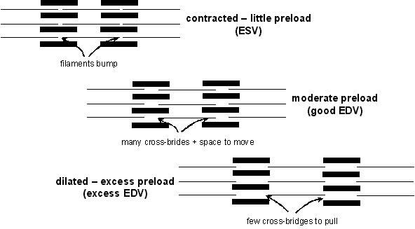
(1) preload = stretch of
cardiac muscle (p. 426, 427)
(sketch)
- can be changed by dilation
Normal
cardiac muscle, medium power microscopic *
Heart,
dilated cardiomyopathy, gross [XRAY]
Heart,
dilated cardiomyopathy, gross

(2) contractility
= strength based on chemistry (O2, ions, hormones, pH, etc.)
- can be changed by hypertrophy
5.
Hypertrophy, heart, gross
Heart,
hypertension with left ventricular hypertrophy, gross
Heart,
cardiomyopathy, microscopic *
(3) afterload = resistance
to emptying from chamber diameter (ventricular radius), BP in aorta or
pulmonary artery, wall thickness and stiffness (p.
427 )
- can be changed by dilation and hypertrophy
(sketch)
Heart,
dilated cardiomyopathy, gross [XRAY]
Heart,
dilated cardiomyopathy, gross
5.
Hypertrophy, heart, gross
Heart,
hypertension with left ventricular hypertrophy, gross
Chapters 28-29
PRELOAD PRELOAD PRELOAD PRELOAD PRELOAD
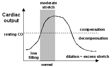
Within normal limits, a
little increase in stretch = a little increase in preload, which is a GOOD thing
incr. stretch -> incr.
preload -> incr.
strength -> incr.
SV -> incr.
CO -> incr.
BP -> incr.
blood flow
(i.e., normal heart adjusts preload to increase or decrease CO as needed {e.g.,
exercise, rest})
With excess blood in the heart (i.e.,
excess End Diastolic Volume {EDV}) -> excess
stretch -> excess preload, which is a BAD thing
incr. incr. stretch -> incr.
incr. preload -> decr.
strength -> decr.
SV -> decr.
CO -> decr.
BP -> decr.
blood
flow (trouble area)
![]()
Compensation - a little increase in stretch ->
a little increase in preload -> a GOOD thing
incr. needed flow -> incr. venous return -> incr. stretch -> incr. preload -> incr. blood flow
ABNORMAL
Above normal limits, - excess stretch
-> excess preload ->
a BAD thing
e.g.,
*(1) incr. incr. DILATION from ischemia = passive congestion
(2) very high BP
(3) injured heart cells
(4) arrhythmias
(5) bad valves
(6) weak cells from low contractility
incr. incr. EDV -> incr. incr. stretch -> incr. incr. preload -> decr. strength -> decr. SV -> decr. CO -> decr. BP -> decr. blood flow (trouble area)
acutely, decr.
SV
from one beat ->incr.
incr. ESV for next beat ->
incr. incr. EDV ->
incr. incr. incr. stretch ->
incr. incr. incr. preload -> decr.
decr. blood flow ->
etc., etc.
(i.e.,
one weak stroke -> an even weaker next stroke ->
an even weaker next stroke -> etc.)
Decompensation
chronically, long term incr. EDV or long term ischemia -> permanent DILATION ->incr. incr. incr. stretch -> incr. incr. incr. preload ->decr. decr. decr. blood flow -> etc., etc.
Normal
cardiac muscle, medium power microscopic *
Heart,
dilated cardiomyopathy, gross [XRAY]
Heart,
dilated cardiomyopathy, gross
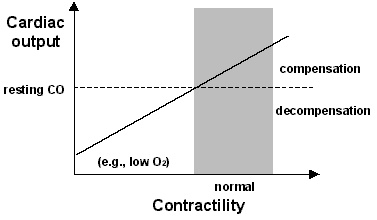
CONTRACTILITY CONTRACTILITY CONTRACTILITY
(p.426)
NORMAL
Compensation –
an increase in contractility (strength) is always a
GOOD thing because contractility = strength
(e.g., by vasodilation of coronary arteries)
incr. needed flow
or mild ischemia from work -> incr.
coronary
vasodilation -> incr.
contractility -> incr.
blood
flow
(i.e., normal heart contractility to increase or decrease CO as needed {e.g.,
exercise, rest})
ABNORMAL
acutely, - a decrease in contractility (strength) is always a BAD thing
decr. O2 (ex. respiratory problems, narrow vessels from thrombus) -> decr. contractility -> decr. blood flow
chronically, (with atherosclerosis)
-
a decrease in contractility (strength) is always a BAD thing
incr.
needed flow
or mild ischemia from work ->
no coronary vasodilation -> decr.
contractility
(ex. O2,
incr.
CO2,
pH) -> decr.
blood
flow -> etc., etc.
Decompensation – from hypertrophy, which raises heart O2 use above blood supply to heart muscle
chronically, a
decrease in contractililty (strength) is a BAD thing
incr. incr. overwork (ex. high BP, bad valves) -> incr. incr. cardiac HYPERTROPHY -> incr. incr. O2 demand -> decr. decr. contractility -> decr. decr. blood flow -> etc., etc.
5.
Hypertrophy, heart, gross
Heart,
hypertension with left ventricular hypertrophy, gross
Heart,
cardiomyopathy, microscopic *
To make matters worse,
excess
decreased contractility +
excess preload is a VERY BAD thing
decr. contractility -> decr. SV -> incr. incr. ESV -> incr. incr. EDV -> incr. incr. preload -> decr. decr. blood flow -> etc., etc.
i.e.,
one weak beat -> an even weaker next beat ->
an even weaker next beat -> etc.
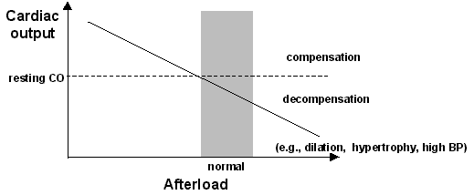
incr. afterload -> decr. SV -> decr. CO -> decr. BP -> decr. blood flow
NORMAL
Compensation
- preload and contractility overpower
(compensate for) afterload
(i.e., normal heart adjusts preload and
contractility to compensate for normal increase or decrease in afterload)
incr.
ventricular radius -> incr.
afterload but also incr. preload ->
incr.
SV -> incr.
CO -> incr.
BP -> incr.
blood flow
incr.
BP -> incr.
afterload but also incr.
contractility from coronary vasodilation
-> incr.
SV -> incr. CO -> incr.
BP -> incr.
blood flow
ABNORMAL
acutely,
- excess preload is a BAD thing, which also increases afterload ->
a VERY BAD thing
- decreased contractility is a bad thing, which also increases
afterload -> a VERY BAD thing
incr.
incr. EDV from excess preload or bad contractility (DILATION) -> incr.
incr. afterload incr. incr. incr.
preload -> decr.
SV -> decr.
CO -> decr.
BP -> decr.
blood flow and incr. incr. incr.
ESV -> incr.
incr. incr. EDV for next beat -> incr.
incr. incr. afterload and incr. incr.
incr. preload ->
etc. etc.
i.e.,
one weak beat -> an even weaker next beat ->
an even weaker next beat -> etc.
Decompensation
-
excess preload is a BAD thing, which also increases afterload ->
a VERY BAD thing
- decreased contractility is a bad thing,
which also increases
afterload -> a VERY BAD thing
-
excess preload and decreased contractility are BAD things,
which also increase afterload -> a VERY VERY BAD thing
chronically, DILATION from long term ischemia or HYPERTROPHY
from long term overwork -> (sketch)
incr. incr.
afterload with incr. incr.
preload and decr. decr. contractility ->
decr.
SV -> decr.
CO -> decr.
BP -> decr.
blood flow
i.e., one weak
beat -> an even weaker next beat ->
an even weaker next beat -> etc.
#################################################################
NOTE:
– in summary:
Preload
incr.
preload -> slightly increased stretch ->
incr.
strength -> incr.
SV -> etc., etc. (GOOD)
incr.
incr. preload -> excess stretch -> decr.
strength -> decr.
SV -> etc., etc. (from dilation) (BAD)
Contractility
incr.
contractility -> always incr.
strength -> incr.
SV ->
etc., etc. (GOOD)
decr.
contractility -> always decr.
strength -> decr.
SV ->
etc., etc. (from hypertrophy) (BAD)
Afterload
decr.
afterload -> always decr.
resistance -> incr.
SV ->
etc., etc. (GOOD)
incr.
afterload -> always incr.
resistance -> decr.
SV ->
etc., etc. (BAD)
(from either dilation or
hypertrophy) (BAD)
Therefore,
dilation
+ hypertrophy ->
excess preload + decreased contractility + increased afterload
->
VERY VERY BAD
Heart,
dilated cardiomyopathy, gross [XRAY]
Heart,
dilated cardiomyopathy, gross
5.
Hypertrophy, heart, gross
Heart,
hypertension with left ventricular hypertrophy, gross
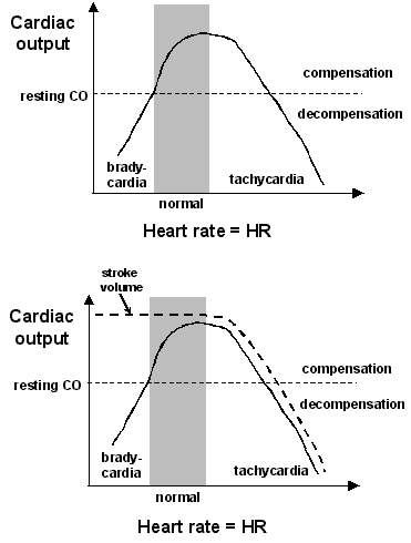
Variations in heart rate = incr. HR or decr. HR
CO = SV
x HR
CO = cardiac output = volume/minute
SV = stroke volume = volume/beat
HR = heart rate = beats/minute
normal = compensation
incr.
HR ->incr.
CO ->incr.
BP ->incr.
blood flow
decr.
HR -> decr.
CO -> decr.
BP -> decr.
blood flow
(i.e., normal heart adjusts HR to increase or decrease CO as needed {e.g.,
exercise, rest})
abnormal = decompensation
(1, 2, & 3)
incr.
incr. incr. HR -> decr.
CO -> decr.
BP -> decr.
blood flow
incr. incr.
HR = tachycardia > 100 BPM at rest
1. incr.
incr. incr. HR -> decr.
filling time ->
decr.
decr. EDV (decr. decr. preload)
-> ??
2. incr.
incr. incr. HR -> incr.
incr. O2 demand -> decr.
contractility -> ??
3. incr.
incr. incr. HR -> decr.
decr. diastole -> incr.
vessel closure ->
decr.
blood flow -> decr.
O2 supply -> decr.
contractility -> ??
Therefore: incr. incr. incr. HR -> decr. decr. preload & decr. contractility -> decr.decr. decr. SV -> decr. decr. CO
also: decr. decr. decr. HR -> decr. decr. CO
decr. decr. decr. HR = bradycardia < 60 BPM
Definitions
ejection fraction = fraction
ejected per beat
normal = 2/3
abnormal (weak beat) -> decr. decr. ejection fraction -> incr. incr. ESV -> incr. incr. EDV -> incr. incr. preload
ventricular tension = diastolic BP
ventricular radius = "width of ventricle" (sketch)
- abnormally
incr.
incr. ventricular tension = incr.
incr. afterload -> decr.
SV -> decr.
CO
incr. incr. ventricular radius (dilation) = incr. incr. preload & incr. incr. afterload -> decr. SV -> decr. CO
cardiac reserve = incr.
CO when needed (5X)
abnormalities -> decr.
cardiac reserve ->
limited
incr.
CO when incr.
incr. CO is needed
Cardiac oxygen - supply vs. demand (p.
461)
- constant pumping -> constant
O2 demand
- O2 for energy
- if O2 supply less
than O2 demand
- low O2
->
low energy -> decr. CO
- low O2
->
incr.
lactic acid -> cell injury/cell death
Factors affecting O2 demand (p.
460) (1, 2, 3, &4)
1. heart rate - incr.
HR -> incr.
demand
2. contractile force - incr.
force -> incr.
demand
3. muscle mass - incr.
mass (hypertrophy) -> incr.
demand
4. ventricular wall tension =
afterload
- incr. afterload -> incr.
demand
-from ventricular
radius & BP
Normal:
incr. O2
demand -> coronary
vasodilation -> incr.
O2 supply
Abnormal:
incr. O2
demand -> low O2 supply = ischemia
Factors in left ventricle (1, 2, & 3)
1. large mass = high O2
demand
2. large contractile force = high
O2 demand
3. intermittent flow = low O2
supply
- systole
-> vessel compression
Coronary atherosclerosis
Risk factors (see above)
Development of atheroma(see above) (p. 462)
Atheromas -> decr.
blood flow (see above)
-narrowness
(p.
103), roughness (p. 102), stiffness
Effects from ischemia (O2 supply
< O2 demand) (1, 2, & 3)
1. weak contraction (low
contractility)
2. dilation (distention) -> incr.
incr. preload and
incr. incr.
afterload
3. wall rigidity -> incr.
incr. afterload -> decr. decr.
SV
1. + 2. + 3. -> decr. decr. CO -> systemic ischemia
THEREFORE: cardiac ischemia -> 1, 2, & 3
1. initial
decr.
decr.
CO from ischemia +
2. continued
decr.
decr.
CO from cardiac decomp. +
3. problems
from decr. respiratory function
S&S from coronary ischemia (1, 2, & 3)
1. angina
(sublethal injury reversible)
2. T-wave
inversion (p. 467)
(sketch)
3. ST segment
depression
(p. 467) (sketch)
Effects from myocardial infarction (M.I.)
- same as ischemia but WORSE!!
- more severe + longer
lasting (cell death)
Therefore: (1, 2, & 3)
1. decr.
decr. decr.
CO +
2. incr.
incr. incr. decomp. +
3. decr.
decr. decr. respiratory function
S&S from myocardial infarction (M.I.) (1, 2, & 3)
1. lasting pain (lethal injury irreversible)
- also
nausea, vomiting, panic, sweat
2. EKG changes (p.
471)
- inverted
T-wave (sketch)
- ST-segment
elevation (sketch)
3. cardiac enzymes in blood
1 + 2 + 3 = diagnostic triad
Compensatory responses (1, 2, & 3)
1. immediately -> incr.
sympathetic activity
- incr.
rate, incr. force, incr.
vasoconstriction
- decompensation
from incr. incr. O2 demand
2. later -> incr.
hormones
- norepinephrine
incr.
CO, incr. BP , mineralocorticoids
incr.
BP
- decompensation
from incr. incr. O2 demand
3. long term ->
cardiac hypertrophy
-decompensation
from incr. incr. O2 demand,
incr.
incr.afterload
Healing of infarct (2nd intention) -> scar
Complications from M.I. (1 - 10)
1. congestive heart failure
- from
gradual
hypertrophy + dilation (sketch)
- decr.
decr. CO & pulmonary congestion &
systemic congestion
Heart,
dilated cardiomyopathy, gross [XRAY]
Heart,
dilated cardiomyopathy, gross
5.
Hypertrophy, heart, gross
Heart,
cardiomyopathy, microscopic
14.
Remote myocardial infarction, gross
2. cardiogenic shock
-from rapid
dilation
- decr.
decr. CO & pulmonary congestion (&
systemic congestion)
3. papillary muscle dysfunction (p.
472) (sketch)
- valve
allows back flow of blood
- overworked heart & systemic ischemia &
pulmonary congestion
4. ventricular septal defect (p.
472) (sketch)
-blood
from left ventricle to right ventricle
- decr.
decr. CO & overworked heart &
pulmonary congestion
5. cardiac rupture (p.
473) (sketch)
- blood
fills pericardial cavity
- cardiac tamponade
- prevents
filling during diastole -> decr.
decr. CO + pulmonary congestion
6. ventricular aneurysm
(outpocketing) (p. 473)
(sketch)
- CHF + emboli + arrhythmias
15.
Left ventricular aneurysm, gross
7. thromboembolism
(sketch)
- roughness
-> emboli -> strokes, etc.
(8. pericarditis)
(9. Dressler's syndrome)
10. arrhythmias (pp.
476)
- most
common complication
- abnormal
rates (tachy., brady.)
- abnormal
sites of initiation
- ectopic beats, escape beats, (PVC) premature
ventricular contraction (sketch)
- abnormal
conduction -> decr.
decr. CO
- heart blocks (sketch)
- 1st degree, 2nd degree, 3rd degree
- bundle branch block
Treatment strategies (1, 2, 3, & 4)
1. incr.
contractility (incr. O2
supply, meds.)
2. decr.
preload & decr. afterload (decr.
O2 demand)
e.g., decr. fluids, decr.
salt, decr. BP,
3. decr.
workload (decr. O2 demand)
e.g., rest, mechanical pumps
4. surgery
e.g., bypass, valves, transplant, repair defects
Heart valve operation
- direction of blood flow opens
or closes
- smooth
flexible movable sheets
- endothelium
on connective
1. allows forward flow
(sketch)
2. prevents back flow
(sketch)
Causes of valvular heart disease (1 -
5)
1. rheumatic heart disease (most
common)
- Group
A B-hemolytic strep. (sketch)
|
repeated/prolonged rheumatic fever
yields |
||
|
repeated/chronic valve inflammation (?autoimmune?)
yields |
||
|
scar formation in valve
(pp. 489-490) yields |
||
|
1. fused cusps
(adhesions) |
2. stiff cusps | 3. shrunken cusps |
|
1. + 2. ->
stenosis (poor opening) (pp. 491, 494, 496) yields |
2. + 3. ->
regurgitation (insufficiency) (incomplete closing) (pp. 493, 494, 495) yields |
|
|
chronic overworked chamber pushing blood through
valve
yields |
||
|
hypertrophy + dilation of chamber
yields |
||
| CHF | ||
(sketch)
2. bacterial endocarditis(p.
99)
infection
on valve -> inflamed valve
-> scarred valve -> ??
3. damaged papillary muscle(p.
472) (sketch)
M.I. ->
damaged papillary valve -> valve flips backward -> regurgitation
-> ??
4. congenital defect
(sketch)
defect
-> stenosis &/or regurgitation-> ??
5. autoimmunity ("in born
error")
immune
attack on valve -> inflammation ->
scar on valve -> ??
Mitral Stenosis (see Course Booklet p. 36)
Heart chamber that pumps blood through the diseased valve is affected most (pp. 491-496) (sketch)
Heart,
dilated cardiomyopathy, gross [XRAY]
Heart,
dilated cardiomyopathy, gross
5.
Hypertrophy, heart, gross
Heart,
cardiomyopathy, microscopic
Mitral Stenosis (sketch)
|
|
|
|
|
|
|
|
|
|
|
|
|
|
|
|
|
|
|
|
|
|
|
|
|
|
|
|
|
|
|
|
|
|
|
|
|
|
|
|
|
|
|
|
|
|
|
|
|
|
1. decr.
decr. gas exchange
-> dyspnea
2. incr. incr. risk of infection 3. pulmonary fibrosis |
|
|
|
|
|
|
|
|
|
|
|
|
*****************************************************************
WITH NO TREATMENT -> worsening of all
above aspects ->
heart failure &\or respiratory failure
Dangers from pulmonary edema
immediate: thick membrane
+ filled alveoli -> decr.
gas exchange (sketch)
1.
Normal lungs, gross
2.
Normal lung, cross section, gross
4.
Normal lung, microscopic*
Note the thin membranes and open air spaces
5.
Lung, edema, microscopic*
This is at high magnification, note the thick membranes and filled air
spaces
6.
Lung, passive congestion, microscopic*
This is at high magnification, note the thick membranes and filled air
spaces
later: incr.
fluids -> incr.
risk of infection -> decr.
gas exchange (sketch)
17.
Lung, bronchopneumonia, low power microscopic*
chronic:
incr.
fluid -> pulmonary fibrosis ->
decr.
gas exchange
(sketch)
115.
Lung, interstitial fibrosis, microscopic*
Chronic pulmonary edema can cause pulmonary fibrosis.
Degree of damage from atherosclerosis
depends upon (1, 2, &
3)
1. severity
2. tissue
demand
3. amount
of collaterals
Reasons for S&S from ischemia
- from chemical imbalance (decr.
O2, incr. CO2,
etc.)
- intermittent
claudication
- pain
at rest
- necrosis
- atrophy
- cyanosis
- from low blood flow into area
- weak
pulse
- pallor
- coolness
- from pulse wave hitting blockage
- strong
pulse above lesion
1.
Normal lungs, gross
2.
Normal lung, cross section, gross
4.
Normal lung, microscopic*
5.
Lung, edema, microscopic*
6.
Lung, passive congestion, microscopic*
Foot
with previous healed transmetatarsal amputation and recent ulcer, gross.
Gangrenous
necrosis and ulceration, lower extremity, gross.
Aneurysm - outpocketing of vessel
(sketch)
22.
Aorta, atherosclerotic aneurysm, gross
causes (1, 2, 3, &
4)
1. atherosclerosis
2. trauma
3. congenital
defect
4. syphilis
effects (1, 2, &
3) (sketch)
1. thrombus
2. pressure
on nearby structure
3. hemorrhage
Venous thrombosis
causes (1, 2, &
3)
1. roughness
2. slow
flow
3. chemical
imbalance
Renal
vein thrombosis, gross
consequences - block flow
1. at site
of formation (sketch)
2. embolize
to other sites (sketch)
e.g. pulmonary embolus (p. 617)
27.
GIF animation of pulmonary thromboembolus, diagram
69.
Lung, pulmonary thromboembolus, gross
70.
Lung, pulmonary thromboembolus, gross
73.
Lung, pulmonary thromboembolus, gross
74.
Lung, pulmonary thromboembolus, low power microscopic*
27.
GIF animation of pulmonary thromboembolus, diagram
28.
Pulmonary thromboembolus, gross
29.
Pulmonary thromboembolus, gross
30.
Pulmonary thromboembolus, gross
31.
Pulmonary thromboembolus, microscopic *
32.
Pulmonary thromboembolus, microscopic *
78.
Lung, remote organized pulmonary artery embolus, gross
Varicose Veins
(sketch)
causes
(1 - 4)
1. gravity - standing still
2. compressed veins
- posture, tight clothing, pregnancy, tumor
3. back pressure
- coughing, straining with defecation, constipation
4. blocked drainage
- cirrhosis, heart failure
pathogenesis (sketch)
- chronic forced dilation -> permanent
distention (p. 547)
complications
- ischemia, edema, thrombosis, infarction, infection, hemorrhage, pain,
cosmetic
1.
Normal esophagus, gross
2.
Normal esophagus, low power microscopic*
12.
Esophageal varices, gross
13.
Esophageal varices, gross
14.
Esophageal varices, with sclerotherapy, gross
118.
Prolapsed true hemorrhoids, gross
21.
Esophageal varices with portal hypertension, gross
22.
Splenomegaly with portal hypertension, gross
20.
Caput medusae of skin with portal hypertension, gross
Shock
causes
- hemorrhage, heart failure, pulmonary embolus, trauma, severe infection,
stroke, etc.
areas
affected most
-
first
1. brain (high O2 demand)
2. heart (high O2 demand)
3. lungs (pulmonary edema)
-
then
4. kidney (decr. decr.
BP -> decr.
decr. filtration)
© Copyright 2002 - Augustine G. DiGiovanna - All rights reserved. All rights reserved. Except as permitted under the United states Copyright Act of 1976, no part of this page may be reproduced or distributed in any form or by any means, or stored in a data base or retrieval system, without the prior written permission of Augustine G. DiGiovanna.