Biology 334
Pathophysiology
Dr. D.'s Overhead Lecture Notes
Section 3 - REPLACE PAGE NUMBERS WITH PAGES FROM SIXTH EDITION3
Do Not Print This Page
These notes are in your course booklet and in Sec 03-w.htm.
If you click
on a link to a picture, click on "back" to return to these notes.
Nervous System Lecture Notes
Return to
Notes Index
Goals of neurological examination
1. determine existence of neurological
problem
2. determine location of problem
Cerebrovasular disease
- most common cause of neurological disease
CNS (brain) O2 needs
Approx. 2% of body weight / 20% of O2
needs
- high and continuous
O2 demand
- decr.
O2 supply -> immediate cell injury -> immediate
neurological malfunction (reversible)
- prolonged decr.
O2 -> cell death -> prolonged neurological deficit
- no neuron reproduction/replacement
- possible compensation
by remaining neurons
Brain blood supply = brain O2 supply
(pp. 780, 781, 852, 853, 855)
Cerebral
arterial supply, angiogram
8.
Cerebral arterial circulation and aneurysms, diagram
- depends most on cardiac output, not
systemic BP - "me first"
- regulated by self regulating vessels
1. based on
O2, CO2, pH
decr.
O2/incr CO2/decr.
pH -> vasodilation -> ??
incr
O2/decr. CO2/incr
pH -> vasoconstriction -> ??
2. based on
cerebral BP
decr.
cerebral BP -> vasodilation -> ??
incr.
cerebral BP -> vasoconstriction -> ??
- protects against edema
NOTE:
opposite systemic vessels
- assures brain
function by shifting blood to or from brain
- influenced by intracranial pressure
Increased
Intracranial Pressure = IIP
- not a regulatory
process
incr.
intracranial pressure -> vessel compression -> decr.
blood flow -> ??
- causes of
IIP = incr. volume in skull (sketch)
- edema, hemorrhage, tumor, incr.
CSF
Stroke = cerebrovascular accident = CVA
causes - usually from atherosclerosis
Normal
coronary artery, microscopic *
Mild
coronary atherosclerosis, microscopic *
Severe
calcific coronary atherosclerosis, microscopic *
Aorta,
lipid streaks, gross
Aorta,
lipid streaks, gross
Aortas
with mild, moderate, and severe atherosclerosis, gross
Aorta,
atheromatous plaque, medium power microscopic *
Aorta,
atheromatous plaque, high power microscopic *
Aorta,
atheromatous plaque, high power microscopic *
Aorta,
atherosclerotic aneurysm, gross
1. thrombus (p.
853) = most common cause (sketch)
- atheroma ->
thrombus -> blockage -> ischemia
34.
Internal carotid artery, thrombosis, recent, gross [ANGIO]
This image shows blockage of an artery to the brain.
31.
Cerebrum, coronal section, acute cerebral infarct, gross
This image shows infarction from ischemia.
39.
Cerebrum, acute infarction, microscopic *
This image shows infarction from ischemia and the development of edema
from the inflammatory response.
2. embolus - usually from atherosclerosis
(sketch)
e.g., M.I. -> thomboembolus
e.g., carotids -> thromboembolus, plaque
- embolus moves to
brain -> blockage in brain -> ischemia
35.
Cerebral artery, thromboembolus, microscopic *
This image shows blockage of an artery to the brain.
38.
Cerebrum, coronal section, hemorrhagic infarct, from arterial embolus,
gross
This image shows infarction from ischemia. There is also slight hemorrhaging
from the necrotic region.
37.
Cerebrum, coronal section, hemorrhagic infarct, from arterial embolus,
gross
This image shows infarction from ischemia followed by significant hemorrhaging
in the necrotic region. Note the development of edema from the inflammatory
response.
Liquefactive
necrosis, cerebral infarction, gross
This image and the next seven images show the liquefaction of infarcted
brain regions.
Liquefactive
necrosis, cerebral infarction, gross
Liquefactive
necrosis, cerebral infarction, microscopic *
44.
Cerebrum, remote infarct, microscopic *
Liquefactive
necrosis, cerebral infarction, microscopic *
29.
Pons, lacunar infarct, gross
30.
Lacunar infarct, microscopic *
45.
Cerebrum, coronal section, remote infarct, gross
3. hypertensive hemorrhage
- hemorrhage
-> injury from Hb and volume of blood
- i.e., chemical and physical damage from blood
2.
Basal ganglia hemorrhage with hypertension, gross [CT]
3.
Basal ganglia hemorrhage with hypertension, gross [CT]
- hypertensive hemorrhage usually -> more hemorrhage the ruptured aneurysm
- effects from
hypertensive hemorrhage (p. 96) (sketch)
1. ischemia from local decr. BP
2. spurting blood hitting neurons
3. hematoma -> local pressure on neurons
4. hematoma -> IIP -> pressure on all neurons
5. Hb injures neurons (chemical damage)
6. injury -> inflammation -> edema -> IIP -> incr.
neuron injury
47.
Cerebrum, coronal section, edema, gross
This image and the next five images show how edema from ischemic strokes
and hemorrhagic strokes can cause additional brain injury due to pressure,
shifting, and herniation.
52.
Pons, Duret hemorrhages, gross
27.
Pons, Duret hemorrhages, gross
7. IIP (hematoma, -> vessel compression -> diffuse ischemia
8. hematoma -> brain shifting -> injury to neurons and vessels
9. IIP -> herniation through skull openings -> injury to neurons
49.
Cerebrum, uncal herniation from edema, gross
50.
Cerebrum, uncal herniation from edema, gross
51.
Cerebellum, tonsillar herniation from edema, gross
52.
Pons, Duret hemorrhages, gross
27.
Pons, Duret hemorrhages, gross
4. aneurysm - atherosclerosis,
congenital, trauma (p. 855)
- aneurysm ->
hemorrhage -> injury from Hb and volume of blood
- i.e., chemical and physical damage from blood
8.
Cerebral arterial circulation and aneurysms, diagram
10.
Circle of Willis with multiple berry aneurysms, gross
This image and the next four images show "berry" aneurysms, so names because
they appear like berries attached to arteries. Aneurysms can lead to strokes
by being sites of thrombus formation and by hemorrhaging.
11.
Berry aneurysm, sectioned, gross
9.
Ruptured intracranial berry aneurysm, gross
12.
Subarachnoid hemorrhage, base of brain, from ruptured berry aneurysm, gross
13.
Subarachnoid hemorrhage from ruptured berry aneurysm, gross
18.
Vascular malformation, microscopic [CT] *
Aneurysms like these within the brain tissues can cause hemorrhagic strokes
just as berry aneurysms do.
17.
Intracerebral and intraventricular hemorrhage from ruptured vascular malformation,
gross
Here is an example of a hemorrhaging stroke from an aneurysms within the
brain.
Effects from ischemia
ischemia -> decr.
O2/incr. CO2/decr.
pH/decr. nutrients/incr.
wastes -> cell injury/cell death -> brain malfunction
consequences vary based on;
1. regions affected
(area of specialization)
2. amount of injury
a. partial versus complete ischemia
b. rate of onset
- thrombus versus embolus
c. duration
- vasodilation -> incr. blood flow
- dissolving the thrombus -> incr.
blood flow
3. rate of recovery of neurons
Types of strokes based on timing
1. TIA = transient ischemic
attack
- all S&S
gone by 24 hours (may recur)
- long term =
no S&S remain (transient)
2. RIND = residual ischemic neurological
deficit
- S&S last
more than 24 hours
- long term =
some S&S improve quickly and some S&S improve slowly or not at
all (residual)
NOTE: if some
S&S develop later = "progressive stroke" = "stroke in evolution"
e.g., more emboli forming
3. completed stroke
- almost all
S&S develop quickly
- long term =
little or no improvement in S&S
Causes of TIA, RIND, completed strokes
thrombus usually -> TIA (but may -> RIND or
completed stroke)
embolus usually -> RIND (but may -> TIA or completed
stroke)
aneurysm usually -> RIND (but may -> TIA or
completed stroke)
hypertensive hemorrhage usually -> completed
stroke (but may -> TIA or RIND)
S&S (p. 851+
but ignore specific vessels)
Reasons for different S&S
1. regions affected
- have different
functions
2. amount of injury in each area
- mild to severe
3. rate of recovery of neurons
- fast, slow,
none (reversible versus irreversible damage)
Epilepsy
- second to CVA as CNS disorder in adults
- definition
- excessive
- uncontrolled
- synchronous
- paroxysmal
- local
- discharge
- cerebral neurons
(usually in the cortex)
- causes = other cerebral pathologies
e.g., stroke,
trauma, meningitis, alcoholism
Epilepsy may develop as a new disease or it may develop as a complication
from almost any brain pathology.
15.
Brain, parietal lobe, vascular malformation, gross [ANGIO]
18.
Vascular malformation, microscopic [CT] *
60.
Cerebrum, cysticercus cyst, microscopic *
- petit mal = loss of consciousness
- partial or
complete
- brief (moment
to seconds)
- grand mal = loss of consciousness
+ tonic spasms (tension), then clonic spasms (movement)
Multiple sclerosis (MS) = many scars/much
scarring
- cause = idiopathic
- ?second exposure to virus? -> autoimmune
response to CNS myelin
Multiple sclerosis is an autoimmune disease characterized by intermittently
progressive destruction of myelin in the CNS.
- pathogenesis
repeated autoimmune response
yields
repeated myelin injury and inflammation
yields
myelin destruction (-> neuron malfunction) + scar
formation (sclerosis)
yields
repeated in other CNS areas
yields
multiple sclerosis
yields
multiple and progressive CNS
malfunction
83.
Cerebrum, coronal section, multiple sclerosis, gross
84.
Cerebrum, coronal section, multiple sclerosis, gross [MRI]
- prognosis
variable flairs & remissions
(different timing and areas
affected)
yields
variable worsening of conditions
(variable timing and S&S)
Myasthenia gravis (pp.
869, 870) (sketch)
autoimmunity
to neuromuscular junctions
yields
progressive muscle weakness
yields
respiratory failure
Brain trauma (traumatic
injury)
Normal conditions
for brain (pp. 778, 780, 783, 793, 889)
- NOTE: skull (cranium), meninges, brain, CSF, vessels, intracranial pressure
Causes and immediate
effects
1.
penetration or laceration (sketch)
physical objects contact
brain
yields
neuron injury/neuron death
yields
brain malfunction
2. physical force (energy) (sketch)
force (energy) applied to
brain from
(a. blow to head -> shock waves)
(b. rapid penetrating object
-> shock waves)
yields
shock waves
yields
neuron injury/neuron death
yields
brain malfunction
26.
Brain, coronal section, contusions and lacerations with blunt force trauma,
gross
3.
acceleration/deceleration of head (sketch)
rapid start or stop of head
movement
yields
a. brain and skull collide
(sketch)
b. brain rebounds ->
brain and skull collide on other side(contre coup injury)
(sketch)
c. brain shifts in skull
-> scraping/tearing of vessels/nerves
yields
neuron injury/neuron death
yields
brain malfunction
21.
Inferior frontal and temporal lobes, contracoup injury, contusions and
subarachnoid hemorrhage, gross
22.
Inferior frontal lobes, contracoup injury, contusions, gross
23.
Inferior frontal lobes, contracoup injury, contusions, gross
Secondary effects of brain
trauma
1. hemorrhage
outside brain (only physical damage from blood)
6.
Bridging veins from dura, gross
a.
epidural hematoma (pp. 887,
890) (sketch)
- hematoma between dura mater and skull
- usually from artery
- rapid and high pressure bleeding
4.
Epidural hematoma, gross [CT]
b.
subdural hematoma (p. 890)
(sketch)
- hematoma between dura mater and pia mater
- usual from vein
- slow, gradual incr.
pressure
- acute = problems within 24-48 hours
- subacute = problems continue 48 hours - 2 weeks
- chronic = problems for more than 2 weeks
5.
Subdural hematoma, acute, gross [CT]
7.
Subdural hematoma, bilateral, chronic, gross [CT]
59.
Infected subdural hematoma, gross
-
effects from epidural / subdural hematoma
(only physical damage from blood)
- enlarging hematoma -> (sketch)
1. local pressure on neurons ->
2. **IIP -> pressure on all neurons ->
3. **IIP -> vessel compression -> ischemia (p.
885) ->
4. brain shifting -> injury to neurons and vessels ->
5. **IIP -> brain herniation -> injury to neurons ->
yields
neuron injury/neuron death
yields
brain malfunction
2. inflammation
neuron injury/neuron death -> inflammation -> edema ->
IIP ->
1. neuron injury
2. shifting
3. vessel compression
4. herniation
- may include brain cell swelling (hydropic change) -> shifting and IIP
-> etc.
- limited compensation for IIP
- CSF shifts from cranium to scalp &/or spinal cord
(sketch)
3. vessel damage
from trauma
vessel damage -> reduced vessel autoregulation (chemicals, BP) ->
yields
a. inappropriate vasoconstriction -> ischemia ->
b. inappropriate vasodilation -> hyperemia -> IIP ->
yields
neuron injury/neuron death
yields
brain malfunction
Causes of increased intracranial
pressure (IIP)
1. hemorrhage (inside
brain or outside brain)
2. inflammation
3. brain cell swelling
4. tumors
5. excess CSF production
6. inadequate CSF
removal
65.
Cerebrum, coronal section, hydrocephalus, gross
7. passive congestion
(e.g., tumor on vein)
Damage from IIP
1. neuron injury
2. shifting
3. vessel compression
4. herniation
47.
Cerebrum, coronal section, edema, gross
48.
Cerebrum, external view, edema, gross
49.
Cerebrum, uncal herniation from edema, gross
50.
Cerebrum, uncal herniation from edema, gross
52.
Pons, Duret hemorrhages, gross
27.
Pons, Duret hemorrhages, gross
51.
Cerebellum, tonsillar herniation from edema, gross
S&S from brain trauma
- like S&S from stroke
- depends upon
1. regions affected
2. amount of injury
3. rate of neuron recovery
Spinal cord trauma (traumatic
injury)
Normal conditions
for spinal cord (p. 793)
NOTE: vertebrae, adipose, meninges, cord
Causes and immediate
effects
1. penetration or laceration
- physical objects contact cord -> ??
2. excessive flexion/extension/rotation
- bending/twisting -> ??
yields
neuron injury/neuron death
yields
cord malfunction
systemic
effects
1. decr.
voluntary motor function (descending tracts)
2. decr.
conscious sensory function (ascending tracts)
3. decr.
BP from decr.
sympathetic tone (descending tracts and gray matter)
4. decr.
reflexes below site of injury (descending tracts and gray matter) (sketch)
- decr.
brain facilitation of reflexes
- spinal shock
1 & 2 are often permanent
3 & 4 are usually temporary
- types, degree, and duration of effects depends upon (like the
brain)
1. regions affected
2. amount of injury
3. rate of neuron recovery
CNS tumors (neoplasia)
- "primary"
from CNS connective tissue
- neurons do not reproduce
- "metastatic"
from metastasis (e.g., lung, breast)
141.
Cerebrum, coronal section, metastasis, gross
glioma (glioblastoma)
- usually malignant
- most common primary brain cancer
- rapid & invasive -> poor prognosis (death in <1yr. from diagnosis)
134.
Brainstem, sagittal section, glioma, gross
135.
Cerebrum, coronal section, glioma, gross [MRI]
136.
Glioma, low power, microscopic *
133.
Brainstem, external view, glioma, gross
132.
Cerebrum, coronal section, glioma with mass effect, gross [MRI]
138.
Cerebrum, coronal section, glioblastoma multiforme, gross
140.
Gliolbastoma multiforme, high power, microscopic [IPX] *
meningioma
- usually benign
- usually dura mater or arachnoid
- usually slow growing, easily removed by surgery
112.
Meningioma, parasagittal, gross
113.
Meningioma, parasagittal, gross
114.
Meningioma with cerebral compression, gross [MRI]
115.
Meningioma, sphenoid ridge, gross
The meningioma in this image is in the upper left region. It is not in
the upper right region as stated under the image.
116.
Meningioma, resected, gross
120.
Posterior fossa, sagittal section, fourth ventricle with ependymoma, gross
[MRI]
121.
Cerebrum, horizontal section, fourth ventricle with ependymoma, gross
128.
Acoustic nerve schwannoma, gross [MRI]
pathological
effects possible (sketch)
1. local
pressure on neurons -> ??
2. local
pressure on vessels -> ischemia -> ??
3. local
pressure on veins -> IIP from passive congestion & blocked CSF removal
-> ??
65.
Cerebrum, coronal section, hydrocephalus, gross
4. local
pressure on CSF passages -> IIP -> ??
5. weak vessels
-> hemorrhage -> ??
6. general
IIP -> ??
7. competition
for O2 & nutrients (malignant neoplasia) -> ??
yields
neuron injury/neuron death
yields
brain malfunction
classic S&S
from brain tumors
1. headache from pressure on meninges, vessels, cranial nerves
2. vomiting from pressure on medulla
3.
papilledema (swelling of optic disc) from IIP and passive congestion
Alzheimer's disease
For
more details, go to the tutorial on CNS degenerative diseases.
Brain atrophy
Alzheimer's
disease, gross.
Alzheimer's
disease, gross.
Alzheimer's
disease, gross.
Plaques
97.
Cerebrum, Alzheimer's disease, plaque, microscopic, Bielschowsky silver
stain
98.
Cerebrum, Alzheimer's disease, plaque, microscopic, Bielschowsky silver
stain *
100.
Cerebrum, Alzheimer's disease, plaque, microscopic, Bielschowsky silver
stain *
99.
Cerebrum, Alzheimer's disease, plaque, microscopic, Congo red stain
*
Neurofibrillary tangles
101.
Cerebrum, Alzheimer's disease, tangle, microscopic, H&E stain *
Neurofibrillary
tangles, Alzheimer's disease, microscopic
103.
Cerebrum, Alzheimer's disease, tangle, microscopic, Bielschowsky silver
stain *
For more images and other nervous system diseases, go to the SSU network
from on-campus computers at the following URL.
\\Fs_apollo\USER\SCHOOL\HENSON\BIOL\Webpath\334\Web-lists\Web-list-nerv.htm
Respiratory System Lecture Notes
Respiratory System
Purposes for gas exchange
1. obtain O2 - for energy
2. eliminate CO2 - for chemical
balance and to prevent acidosis
3. regulate pH - to remove CO2
to incr. pH or retain CO2
to decr. pH
- compensates
for pH changes from other sources (e.g., food, metabolism)
Note: must occur just fast enough to balance
O2 use, CO2 production, and pH changes
Requirements for proper gas exchange
1. proper ventilation (breathing)
a. open
airways
- normal - open airways and spaces
2.
Normal lung, cross section, gross
4.
Normal lung, microscopic*
- abnormal - filled air spaces
5.
Lung, edema, microscopic*
17.
Lung, bronchopneumonia, low power microscopic*
115.
Lung, interstitial fibrosis, microscopic*
b. proper
pressure changes
c. proper
lung compliance
- elasticity
- surface tension (surfactant)
2. proper perfusion (blood
flow)
a. open
vessels (p. 561)
b. many
capillaries near alveoli (p.
557)
- normal - thin alveolar walls containing capillary networks
2.
Normal lung, cross section, gross
4.
Normal lung, microscopic*
- abnormal - fibrosis replaces the capillary network in the alveolar membrane
115.
Lung, interstitial fibrosis, microscopic*
c. proper heart
function
(p. 559)
shunt unit = low ventilation/good
perfusion
(p. 564)
dead space unit = low perfusion/good
ventilation
(p. 564)
3. proper diffusion
a. large
surface area
b. thin
surface (respiratory membrane)
- normal - large amount
of thin surfaces
2.
Normal lung, cross section, gross
4.
Normal lung, microscopic*
- abnormal - thick membranes
and filled air spaces
29.
Lung, interstitial pneumonitis, microscopic*
5.
Lung, edema, microscopic*
17.
Lung, bronchopneumonia, low power microscopic*
115.
Lung, interstitial fibrosis, microscopic*
Major types of respiratory diseases
1. acute infection
9.
Lung, bronchopneumonia, gross
17.
Lung, bronchopneumonia, low power microscopic*
2. chronic respiratory disease
a. chronic
obstructive
pulmonary disease (COPD) - expiration is main problem
b. restrictive
pulmonary disease - inspiration is main problem
3. cancer
80.
Lung, squamous cell carcinoma, gross [XRAY]
87.
Lung, bronchioloalveolar carcinoma, microscopic*
Results
from inadequate gas exchange
inadequate gas exchange
yields
decr.O2,
incr.
CO2,
decr.
pH
yields
cell injury/cell death
yields
body malfunction (illness/death)
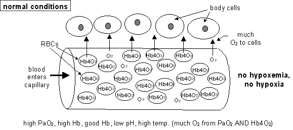
Hypoxemia vs tissue hypoxia
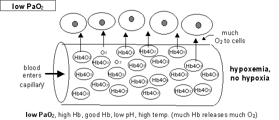
(1) hypoxemia = low dissolved O2 in blood =
low PaO2 = low arterial blood O2
PaO2
= dissolved O2 in arterial blood (from arterial
blood gas analysis)
- measured in mmHg (pressure)
- depends upon proper ventilation only (not
on pH, temp. or Hb)
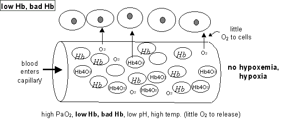
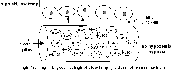
(2) tissue hypoxia = low O2 supply to cells
- most O2 in the blood (95%) is carried bound to Hb as HbO2
-
each Hb can bind up to four molecules of O2 -> actually up
to Hb4O2
(i.e., Hb, HbO2,
Hb2O2, Hb3O2, Hb4O2)
-
therefore, amount of O2 available from HbO2 depends mostly
upon amount of Hb, condition of Hb (e.g., sickle cell anemia, carbon monoxide
poisoning), pH, and temp.
(p. 565)

- good Hb carries much O2 to capillaries near
cells, bad Hb carries little
-
decr. pH, or incr. temp. (all from cell metabolism) -> O2 leaves Hb,
goes to cells
(opposite conditions and effects in lungs to help bind O2 to
Hb)
(To see conditions in lungs, click here.)
- usually only part of
O2 from HbO2 is used
-
unused HbO2 remains in blood unless extra O2 is needed by
cells
- therefore, HbO2
is reservoir for O2 , and can supply more if pH drops or temp. rises
(e.g., high cell metabolism or cell need)
- therefore, since the amount of O2 available for
cells depends on (1) PaO2, (2) amount of Hb, (3) condition of Hb, (4)
pH, and (5) temp. (p. 567), it is possible to have low PaO2
(hypoxemia) and not have hypoxia

- and since the PaO2
supplies only a small portion of the O2 for cells, it is
possible to have a good PaO2 (no
hypoxemia) and still have hypoxia


Therefore, to evaluate hypoxemia and hypoxia, you must know;
1. PaO2 (for hypoxemia = low arterial blood O2)
2. ** amount of Hb in blood
3. ** condition of Hb
4. ** pH
5. ** temperature
(** for hypoxia)
Development of pulmonary edema
(sketch)
1. [decr.
CO -> ] pulmonary congestion
-> incr. pulmonary
BP (>25mmHg/10mmHg)
or
2. decr.
blood proteins -> decr.
COP
or
3. pulmonary inflammation ->incr.
pulmonary BP + decr.
COP |
|
yields
|
|
incr.
fluid leaves capillaries &/or decr.
fluid returns to capillaries
|
|
yields
|
|
incr.
fluid around cells + incr.
fluid in air spaces = pulmonary edema (p.
726)
|
|
|
|
|
|
incr.
fluid around cells
|
&/or
|
incr.
fluid in air spaces
|
|
yields
|
|
yields
|
|
decr.
diffusion
|
|
decr.
ventilation
|
|
yields
yields
|
1. decr. gas
exchange (from thick membranes and filled air spaces)
2. incr. risk
of
infection (from fluids in air spaces)
3. pulmonary fibrosis (from chronic pulmonary edema) |
(sketch)
- normal lung
- much thin membranous surface area and open clear airways and spaces
- normal - much thin membranous
surface area and open clear airways and spaces
2.
Normal lung, cross section, gross
4.
Normal lung, microscopic*
- abnormal lung from pulmonary
edema
(sketch)
- thick membranes
29.
Lung, interstitial pneumonitis, microscopic*
- fluid-filled air spaces
- thick membranes and fluid-filled air spaces
5.
Lung, edema, microscopic*
- infection in fluid-filled air spaces
- infection in fluid-filled air spaces
17.
Lung, bronchopneumonia, low power microscopic*
- fibrosis
- thick membranes and fibrosis
115.
Lung, interstitial fibrosis, microscopic*
Resistances to ventilation
nonelastic resistance
- narrow
airways inhibit flow
- highest
during expiration
- airways collapse during expiration
- overcome
by elastic recoil (passive) or muscles (active, uses O2, makes
CO2)
elastic resistance
- elastic
recoil (from tissue elasticity and surface tension) and tissue stiffness
- highest
during inspiration
- overcome
by surfactant (passive) and muscles (active, uses O2, makes
CO2)
Obstructive disease = incr.
nonelastic resistance
- main problem is expiration
Restrictive disease = incr.
elastic resistance
- main problem is inspiration
Work of breathing = O2
used for ventilation
- usually less than 5% of O2
taken in -> >95% available for body cells
obstructive disease or restrictive disease
yields
incr. resistance
to ventilation
yields
incr. muscle
contraction for ventilation
yields
incr. O2
use = incr. work of
breathing (up to 25% of O2)
yields
decr. O2
available for body cells (only 75%) + incr. CO2
+ decr. pH
See charts for Asthma, Chronic bronchitis, and Emphysema
Asthma
causes
- external
cause = extrinsic asthma
- e.g., dust, pollen, cold air
- internal
cause = intrinsic asthma
- e.g., emotional upset, stress
- either
external or internal cause =mixed
asthma (most common)
pathogenesis (p.
591, 137, 138) (sketch)
|
external irritant &/or internal mechanism
|
|
yields
|
|
histamine release
|
|
yields
(pp. 137, 591)
|
1. mucosal edema
58.
Bronchial asthma, low power microscopic*
2. incr.
mucous secretion
58.
Bronchial asthma, low power microscopic*
59.
Bronchial asthma, high power microscopic*
57.
Bronchial mucus plug with asthma, gross
3. bronchospasms |
|
yields
|
narrow airways
(especially in expiration)
|
|
yields
|
decr. expiration
1.
Normal lungs, gross
55.
Lungs, hyperinflation with status asthmaticus, gross |
|
yields
|
|
decr. ventilation
& incr.work
of breathing (p.145)
|
|
yields
|
decr. O2,
incr.
CO2, decr. pH
(reciprocal with incr.
work of breathing)
(p. 145)
|
|
yields
|
|
cell injury &/or cell death
|
|
yields
|
|
body malfunction
|
S & S = dyspnea,
difficult expiration, wheeze
Chronic bronchitis
causes = smoking,
any air pollution
(pp.
592-594)
pathogenesis
(sketch)
|
chronic air pollution
|
|
yields
|
|
chronic airway inflammation
|
|
yields
(p.594)
|
1. mucosal edema
2. incr. mucous
secretion
3. decr. ciliary
action |
|
yields
(p.594)
|
narrow airways
(especially in expiration)
|
|
yields
|
|
decr. expiration
|
|
yields
|
decr. ventilation
&
incr. work
of breathing
(especially with coughing)
|
|
yields
|
decr. O2,
incr.
CO2, decr.
pH
(reciprocal with incr.
work of breathing)
|
|
yields
|
|
cell injury &/or cell death
|
|
yields
|
|
body malfunction
|
S & S = dyspnea,
chronic cough, incr.
sputum
(p. 553)
Emphysema - Centrilobar emphysema (CLE)
(sketch)
- gross views
2.
Normal lung, cross section, gross
66.
Lung, centrilobular emphysema, gross
- microscopic views
4.
Normal lung, microscopic*
68.
Lung, emphysema, microscopic*
causes = smoking,
any air pollution
(pp. 592-594)
pathogenesis (sketch)
|
chronic air pollution
|
|
yields
|
|
chronic airway inflammation
|
|
yields
|
|
enlarged & weakened bronchi (p.
592)
|
|
yields
yields
|
|
collapsed airways during expiration (p.
594)
|
decr. blood
vessels
|
|
yields
|
yields
|
|
decr. expiration
|
pulmonary hypertension
|
|
yields
|
yields
|
decr. ventilation
&
incr.
work of breathing
(especially with coughing)
|
overworked heart
|
|
yields
|
yields
|
decr. O2,
incr.
CO2,
decr.
pH
(reciprocal with incr. work
of breathing)
|
cor pulmonale
(right-sided heart failure from pulmonary hypertension)
(p. 619)
|
|
yields
|
|
cell injury &/or cell death
|
|
yields
|
|
body malfunction
|
S & S = dyspnea,
cough, incr.
sputum, low FEV1
Emphysema - Panlobar emphysema (PLE)
(sketch)
causes = air
pollution, genes, "aging", ??
(pp. 592-594)
pathogenesis
(sketch)
|
bronchitis, genes, "aging", ??
|
|
yields
|
|
distention & fusion of alveoli
|
|
yields
yields
|
1. weak and collapsing airways
(decr.
expiration -> decr. ventilation)
2. lung rigidity
(decr.
expiration -> decr. ventilation)
3. decr. capillaries
(decr.
perfusion)
(sometimes ->
cor pulmonale)
4. decr. surface
area
(decr.
diffusion) |
blebs
(bubble-like bulge on lung surface)
|
|
yields
|
yields
|
|
(# 1-4 combined)
|
pneumothorax
|
|
yields
|
decr. O2,
incr. CO2, decr. pH
(reciprocal with incr. work
of breathing)
|
|
yields
|
|
cell injury &/or cell death
|
|
yields
|
|
body malfunction
|
S & S = dyspnea,
barrel chest, low FEV1 (p. 553)
Interrelatedness among obstructive diseases
(p. 591)
(sketch)
Complications form COPD
bleb -> pneumothorax
(compression atelectasis = pressure collapses lung) -> decr.
inspiration
(p. 593, 604)
(sketch)
65.
Lungs, bullous emphysema, gross
bulla (large air-filled
space in lung) -> collapsed airway
-> permanent collapse of region served (absorption atelectasis =
removal of air collapses lung) (p. 593) (sketch)
cor pulmonale (sketch)
- right-sided
heart failure from pulmonary hypertension (p. 619)
pneumonia = inflammation
in the lung (sketch)
- pulmonary
edema, incr.
mucus, decr.
clearing -> incr.
risk of infection
pulmonary edema (sketch)
Problems from O2 therapy with
COPD
COPD
yields
chronic
incr.
CO2 from decr.
ventilation
yields
brain uses decr.
O2
(not incr. CO2) to stimulate
ventilation
- therefore
incr.
O2 from O2 therapy
yields
decr.
ventilation even with incr.
CO2
yields
acidosis
yields
etc., etc.
Restrictive respiratory diseases
- main effect is decr.
inspiration with incr.
elastic resistance
extrapulmonary types
- abnormality is outside the pleural cavity
neurological
causes
causes - strokes, drugs, head trauma, high cervical fracture
mechanisms - inhibits stimulation of diaphragm and other respiratory
muscles
anatomic
causes
causes - kyphoscoliosis, trauma, obesity
mechanisms - inhibits movement of thoracic components
intrapulmonary types - abnormality
is inside the pleural cavity
- pleural
effusion = fluid in pleural cavity outside lung (p.
604) (sketch)
causes - congestive heart disease, hypoproteinemia, inflammation
of lung surface, hemothorax (blood in thorax)
126.
Pulmonary atelectasis, gross [XRAY]
127.
Hemothorax, gross
128.
Chylothorax, gross [XRAY]
mechanisms - fluid puts pressure on lung and takes up space
fluid -> pressure on lung
-> partial lung collapse (compression atelectasis) ->
decr. compliance ->
decr.inspiration
->
decr.
ventilation ->
decr.
O2, incr.
CO2, decr.
pH -> cell injury/cell death -> body
malfunction
S & S = dyspnea, altered sounds and motion, x-ray (p.
604)
- pneumothorax
= air in pleural cavity outside lung (sketch)
causes - trauma, bleb bursts, surgery
- closed pneumothorax = trapped air can be absorbed
- open pneumothorax = more air enters with each inspiration
mechanisms - air puts pressure on lung and takes up space
air -> pressure on lung + decr.
pressure changes
-> partial lung collapse
+ decr. compliance + decr.
pressure changes -> decr.
inspiration -> -> ->
-> body malfunction
S&S = dyspnea, altered sounds and motion x-ray
- pneumonia
= pneumonitis = inflammation of the lung
causes and mechanisms
- microorganisms (p. 608)
- bacteria -> exudate blocks airways -> decr.
ventilation (sketch)
- bronchopneumonia
7.
Lung, bronchopneumonia, gross [XRAY]
8.
Lung, bronchopneumonia, gross
18.
Lung, bronchopneumonina, high power microscopic*
20.
Lung, abscessing pneumonia, low power microscopic*
- lobar pneumonia
11.
Lung, lobar pneumonia, gross
12.
Lung, lobar pneumonia, gross
13.
Lung, empyema, gross
- pulmonary abcess
14.
Lung, abscesses, gross
S&S = dyspnea, productive cough, altered sounds, chills,
fever, x-ray
- viruses -> edema -> decr. diffusion (sketch)
29.
Lung, interstitial pneumonitis, microscopic*
S&S = dyspnea, dry cough, fever
- TB and fungi -> necrosis of lung tissue -> (decr.
ventilation, decr. perfusion,
decr.
diffusion) (sketch)
- TB
34.
Lung, tuberculosis with granulomatous inflammation, gross
35.
Lung, tuberculosis with granulomatous inflammation, gross
36.
Lung, granulomatous inflammation and caseation, gross
39.
Lung, Ghon complex with primary tuberculosis, gross
40.
Lung, granulomas, low power microscopic*
41.
Lung, granulomas, low power microscopic*
42.
Lung, granulomas, medium power microscopic*
44.
Lung, M. Tuberculosis, acid fast stain, high power microscopic*
45.
Lung, miliary tuberculosis, gross
46.
Lung, miliary tuberculosis, gross
- fungal infections
47.
Lung, Aspergillus, gross
48.
Lung, fungal granuloma, gross
49.
Lung, extensive fungal granulomas, gross
S&S = dyspnea, productive cough, hemoptysis, x-ray, skin
test
- aspiration of gastric contents -> blockage, inflammation,
necrosis, and abscess of airways and lung
4.
Larynx with aspiration of food, gross
14.
Lung, abscesses, gross
15.
Lung, abscesses, gross
- S&S = dyspnea, choking, cough, fever
- dusts -> chronic inflammation -> scar formation (pulmonary
fibrosis) -> decr.
elasticity (decr. compliance)
(p. 612)
110.
Farmer's lung, scene
111.
Silo filler's disease, scene
101.
Fibrous pleural plaques, gross
102.
Fibrous pleural plaque, low power microscopic*
99.
Asbestos body, microscopic*
100.
Lung, ferruginous bodies, iron stain, microscopic*
103.
Lung, silicotic nodule, low power microscopic*
104.
Lung, anthracosis, microscopic*
105.
Lung, silicosis, polarized light microscopic*
115.
Lung, interstitial fibrosis, microscopic*
116.
Lung, interstitial fibrosis, Trichrome stain, microscopic*
- S&S = dyspnea
- reduced
surfactant
- causes
- hyaline membrane disease (premature birth)
- adult respiratory distress syndrome = ARDS
- mechanisms
- increases surface tension
- decr. surfactant
-> incr.
surface tension -> decr.
compliance
- edema (with ARDS)
note: ARDS has decr. surfactant + edema
118.
Lung, diffuse alveolar damage, gross
119.
Lung, diffuse alveolar damage, microscopic*
- S&S = dyspnea, straining to inspire
Cor pulmonale
causes (p.
619)
- chronic
bronchitis, emphysema, pulmonary embolism, pulmonary fibrosis -> decr.
ventilation -> pulmonary vasoconstriction -> pulmonary hypertension
-> overworked right ventricle ->
dilation and hypertrophy -> right CHF
(cor pulmonale)
(p. 617) (sketch)
- decr.
number/size of vessels ->
blocked flow ->
pulmonary hypertension ->
overworked right ventricle ->
dilation and hypertrophy ->
right CHF (cor pulmonale) (sketch)
respiratory failure
hypoxemic respiratory failure
(p. 624)
- from
problems other than ventilation
- low O2
but normal (or low) CO2
hypercapnic respiratory failure
(p. 624)
- from
decr.
ventilation (+ other problems)
- low O2
+ high CO2
respiratory insufficiency
- decr.
O2 or incr.
CO2 during exercise
respiratory failure
- PaO2
50 mmHg (normal = 85-100)
- PaCO2
50 mmHg (normal = 35-44)
pulmonary malignant neoplasms
- i.e. lung cancer (p.
113 fig. 8-7)
- risk factors (causes)
- for primary
lung cancer - air pollution (smoking)
79.
Lung, squamous cell carcinoma, gross [CT]
80.
Lung, squamous cell carcinoma, gross [XRAY]
81.
Lung, squamous cell carcinoma, gross [XRAY]
85.
Lung, peripheral adenocarcinoma, gross
86.
Lung, bronchioloalveolar carcinoma, gross
87.
Lung, bronchioloalveolar carcinoma, microscopic*
88.
Lung, oat cell carcinoma, gross
89.
Lung, oat cell carcinoma, gross
97.
Lung, mesothelioma, gross
- for metastatic
lung cancer
- all systemic blood goes to heart and then the lungs
94.
Lung, metastatic carcinoma, gross [XRAY]
93.
Lung, metastatic carcinoma, gross [XRAY]
- S&S = dyspnea,
hoarseness, difficulty swallowing, chronic cough, hemoptysis, digital clubbing,
x-ray
- effects (sketch)
- decr.
ventilation (blocks airways)
- decr.
perfusion (blocks/replaces vessels)
- decr.
diffusion (thickens/replaces surface {i.e., respiratory membrane})
Do Not Print This Page
Do Not Print This Page
© Copyright 2002 - Augustine G. DiGiovanna - All rights reserved.
All rights reserved. Except as permitted under the United states Copyright
Act of 1976, no part of this page may be reproduced or distributed in any
form or by any means, or stored in a data base or retrieval system, without
the prior written permission of Augustine G. DiGiovanna.


